|
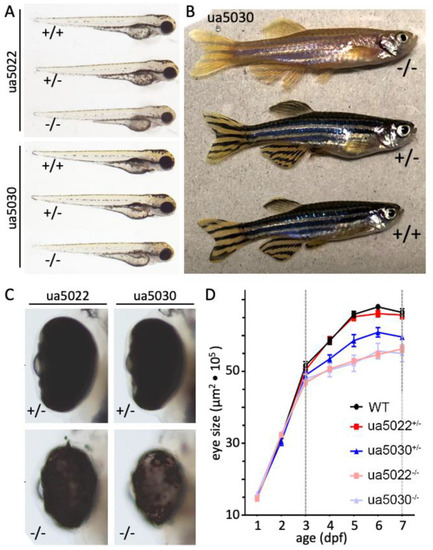
pmela mutants display systemic hypopigmentation and retarded eye size development. (A) Both pmela mutant alleles present with systemic hypopigmentation in homozygotes at 3 days post-fertilization (dpf). (B) Adult homozygous ua5030 zebrafish (−/−, top) display hypopigmentation when compared to heterozygous and wildtype fish. (C,D) Microphthalmia is observed in homozygous mutants from both alleles beginning by 4 dpf (days post-fertilization). Heterozygous pmela+/ua5022 larvae show normal eye size, whereas heterozygous pmela+/ua5030 larvae have reduced eye size. Considering the normal abundance of Pmela in pmela+/ua5030 larvae (Figure 2E), this appears to be a dominant phenotype rather than haploinsufficiency.
|

