Figure 5
- ID
- ZDB-FIG-230916-156
- Publication
- Omar et al., 2023 - Parkinson's disease model in zebrafish using intraperitoneal MPTP injection
- Other Figures
- All Figure Page
- Back to All Figure Page
|
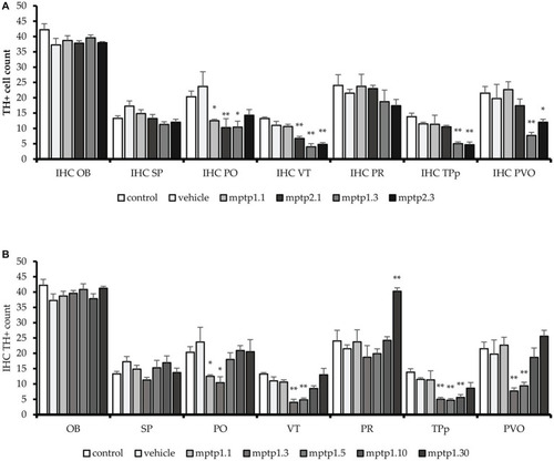
TH+ cells count in different regions of the brain. MPTP in this study has been shown to affect a few areas of dopaminergic neurons, sparing the OB, SP and PR areas. VT and PO area has been seen to be affected the earliest, as early as day 1. Subsequently, other areas were affected. The pattern is similar to other markers tested before, where the lowest counts were noted in MPTP1.3 and MPTP1.5. Day 10 assessment has shown recovery of the cell counts, which mostly return to the same counts as in control on day 10. Although cell counts did not reduce in pretectum following MPTP insult, it is noted that on day 30, the PR cell counts were significantly increased. |

