FIGURE

FIGURE 5

ID
ZDB-FIG-230601-25
Publication
Bai et al., 2023 - Association analysis and functional follow-up identified common variants of JAG1 accounting for risk to biliary atresia
Other Figures
All Figure Page
Back to All Figure Page
FIGURE 5

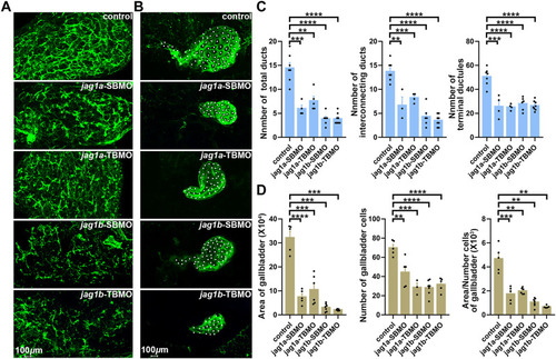
Knockdown of jag1a or jag1b led to biliary defects in zebrafish. (A) Confocal projection on 5dpf control larvae and jag1 morphants stained with cytokeratin 18 showed that the intrahepatic bile ducts of jag1a or jag1b morphants were sparse compared to controls. (B) Confocal projection on 5dpf control larvae and jag1 morphants stained with cytokeratin 18 showed that gallbladders of jag1a or jag1b morphants were smaller or even no compared to those of embryos injected with control MO. Gallbladder cells were noted by white dots. (C) Quantitative analysis of the numbers of total ducts, interconnecting ducts and terminal ductules showed that intrahepatic bile ducts were significantly reduced in both jag1a and jag1b morphants compared with the embryos injected with control MO. (D) Quantitative analysis of the number of gallbladder cells, the measured area of gallbladders and the mean area of individual gallbladder cell showed that both jag1a and jag1b morphants had smaller gallbladders and individual gallbladder cells compared to the embryos injected with control MO. Scale bar = 100 μm. All the data are shown as the mean ± SEM. *p < 0.05; **p < 0.01; ***p < 0.001; ****p < 0.0001. dpf, day post fertilization; SEM, standard error of the mean.

Expression Data

Expression Detail
Antibody Labeling
Phenotype Data
Fish:
Knockdown Reagents:
Observed In:
Stage: Day 5

Phenotype Detail
Acknowledgments
This image is the copyrighted work of the attributed author or publisher, and ZFIN has permission only to display this image to its users. Additional permissions should be obtained from the applicable author or publisher of the image. Full text @ Front Genet