FIGURE

Figure 4

ID
ZDB-FIG-230515-60
Publication
Wilson et al., 2023 - Spinal cords: Symphonies of interneurons across species
Other Figures
All Figure Page
Back to All Figure Page
Figure 4

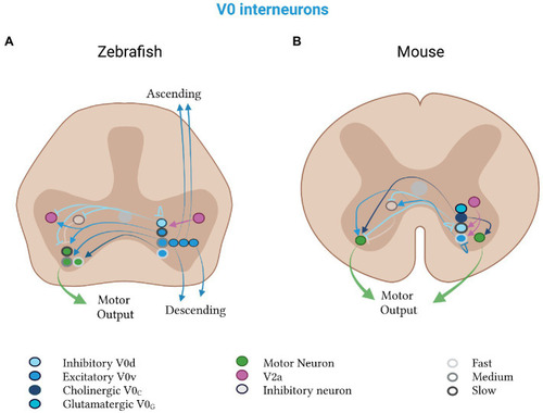
Mixed V0 subtypes in zebrafish and mice. V0d neurons (light blue) inhibit, and V0v neurons (medium blue) excite, contralateral motor neurons (green). (A) Zebrafish V0d neurons receive input from V2a neurons (pink) and project to other V0d, contralateral inhibitory (gray) and ipsilateral excitatory (pink) neurons. The V0v neurons project to V2a neurons and divide into a rhythmic and a non-rhythmic group. The rhythmic group is further split into fast (light gray, outline), medium (medium gray, outline) and slow (dark gray, outline) subtypes, which project to motor neurons of the respective speed class. Excitatory V0v neurons also segregate by projection pattern into ascending, descending and bifurcating subpopulations. (B) Mouse V0d and V0v neurons control slow and fast speeds, respectively. Both classes receive input from V2a neurons. The V0v class additionally projects to contralateral neurons (gray), which inhibit motor neurons. Mouse-specific V0c neurons (dark blue) are cholinergic and project to motor neurons on both sides of the spinal cord. Mouse-specific V0g neurons (turquoise) are glutamatergic and their projection pattern is as of yet unknown.

Expression Data

Expression Detail
Antibody Labeling
Phenotype Data

Phenotype Detail
Acknowledgments
This image is the copyrighted work of the attributed author or publisher, and ZFIN has permission only to display this image to its users. Additional permissions should be obtained from the applicable author or publisher of the image. Full text @ Front. Neural Circuits