FIGURE SUMMARY
Title

Spinal cords: Symphonies of interneurons across species

Authors
Wilson, A.C., Sweeney, L.B.
Source
Full text @ Front. Neural Circuits

A cross-species comparison of the neural basis of vertebrate movement. (A) Cladogram of vertebrate evolution with illustrations of movement patterns for each of the species listed as examples. The lamprey is the most primitive vertebrate and exhibits simple, undulatory swimming; zebrafish display more complex swimming patterns; the frog and salamander use both tail and limbs for movement; reptiles exhibit diagonal limb coordination; and mammals display complex fore−/hindlimb gaits. (B) Cardinal neuron classes that make up the spinal cord circuitry are derived from 11 progenitor domains. Some domains give rise to more than one neuron class, e.g., the p2 domain gives rise to the V2a, V2b, and V2c interneurons. (C) Comparison of interneuron subtypes and projection patterns in the spinal cord of zebrafish versus mice. Colors represent different neuron classes; gray represents neurons without a clear cardinal class identity.

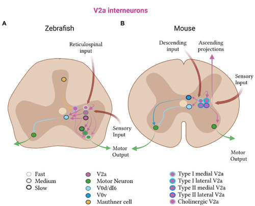
Excitatory V2a subtypes in zebrafish and mice. (A) In zebrafish, the V2a class (pink) is divided into fast (light gray, outline), medium (medium gray, outline) and slow (dark gray, outline) subtypes. The fast subtypes are more dorsal than the slow subtypes. Each subtype receives reticulospinal and sensory input, and projects to the corresponding fast, medium or slow class of motor (green) and V0d (light blue) neurons. Zebrafish also have a cholinergic subclass of V2a neurons, which receive input from Mauthner cells and have bidirectional connections to fast motor neurons. (B) In mice, the V2a population receives descending and sensory input, projects to inhibitory V0d and motor neurons, and has recurrent connections. Mouse V2a neurons subdivide into type I and type II, which further divide into medial and lateral subtypes (shades of blue, outlines). Type I V2a neurons connect to V0 neurons (V0d in light blue and V0v in dark blue); type II V2a neurons have ascending connections.

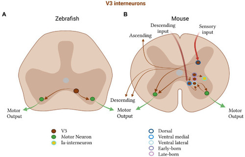
Excitatory V3 subtypes in zebrafish and mice. (A) In zebrafish, V3 neurons (brown) project to motor neurons (green) on both sides of the spinal cord and (B) in mice are divided into a dorsal (dark blue, outline) and ventral class, the latter of which is further subdivided into a medial (medium blue, outline) and lateral (light blue, outline) group. The ventral medial group receives descending commands, forms recurrent connections with itself, and projects to the ventral lateral group. The lateral group projects to motor neurons on the ipsi and contralateral side. Motor neurons project back to V3 neurons in mice. V3 neurons also subdivide by their birth time into an early-(dark purple, outline) and a late-born (light purple, outline) subpopulation, which form either both an descending and ascending, or only a descending, projection, respectively. V3 neurons in mice also project to Ia- interneurons.

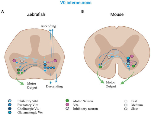
Mixed V0 subtypes in zebrafish and mice. V0d neurons (light blue) inhibit, and V0v neurons (medium blue) excite, contralateral motor neurons (green). (A) Zebrafish V0d neurons receive input from V2a neurons (pink) and project to other V0d, contralateral inhibitory (gray) and ipsilateral excitatory (pink) neurons. The V0v neurons project to V2a neurons and divide into a rhythmic and a non-rhythmic group. The rhythmic group is further split into fast (light gray, outline), medium (medium gray, outline) and slow (dark gray, outline) subtypes, which project to motor neurons of the respective speed class. Excitatory V0v neurons also segregate by projection pattern into ascending, descending and bifurcating subpopulations. (B) Mouse V0d and V0v neurons control slow and fast speeds, respectively. Both classes receive input from V2a neurons. The V0v class additionally projects to contralateral neurons (gray), which inhibit motor neurons. Mouse-specific V0c neurons (dark blue) are cholinergic and project to motor neurons on both sides of the spinal cord. Mouse-specific V0g neurons (turquoise) are glutamatergic and their projection pattern is as of yet unknown.

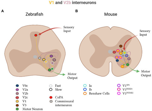
Inhibitory V1 and V2b subtypes in zebrafish and mice. (A) In zebrafish, V1 (yellow) and V2b (brown) divide into fast (light gray, outline) and slow (dark gray, outline) subtypes. V1 neurons: The fast V1 subgroup (yellow, light gray outline) inhibits both slow (green, dark gray outline) and fast (green, light gray outline) motor neurons in addition to slow-type V2a neurons (pink, dark gray outline). The slow V1 subgroup (yellow, dark gray outline) inhibits slow motor neurons (green, dark gray outline). V1 neurons also project to dorsal CoPA neurons (red) which receive sensory input, V2a neurons (pink), V2b neurons (brown), and commissural neurons (gray). V2b neurons: Slow V2b neurons (brown, dark gray outline) inhibit fast motor and other V2b neurons. Fast V2b neurons (brown, light gray outline) inhibit slow motor and other V2b neurons. V2b neurons in zebrafish also project to V2a, V1 and commissural neurons. (B) In mice, V1 neurons and subdivide into Ia-interneurons (light blue, outline), which receive sensory input and inhibit motor output; Renshaw cells (orange outline) which form recurrent connections with motor neurons; and four clades: Sp8 (purple outline), FoxP2 (pink-red outline) and Pou6f2 (pink outline). V1 neurons also receive input from V3 neurons (brown). V2b neurons include Ia- and Ib- (dark blue, outline) interneurons. V2b-derived Ia-interneurons inhibit motor and other V2b neurons. V2b neurons also inhibit V0c neurons. An additional V2c class is present in mice (red-pink) with an unknown projection pattern.

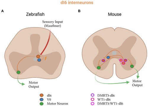
Inhibitory dI6 subtypes in zebrafish and mice. (A) In zebrafish, dI6 neurons (orange) receive input from Mauthner cells, form electrical connections with V0 neurons (blue), and project to contralateral motor neurons (green). (B) In mice, dI6 neurons split into three subclasses: DMRT3- (purple outline), WT1- (pink outline), and DMRT3- and WT1-co-expressing (pink-red outline). WT1-dI6 inhibit contralateral V0 and DMRT3-dI6 neurons, while DMRT3-dI6 inhibit motor neurons on both sides of the spinal cord.

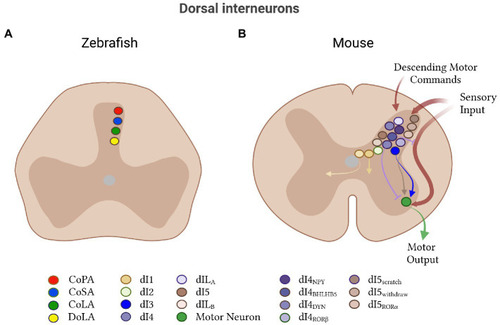
Other dorsal interneurons in zebrafish and mice. (A) In zebrafish, four classes of dorsal interneuron have been identified: glutamatergic commissural primary ascending (CoPA, red), glycinergic commissural secondary ascending (CoSA, dark blue), glycinergic commissural longitudinal ascending (CoLA, green), and glycinergic dorsal longitudinal ascending (DoLA, yellow). CoPA neurons drive touch-mediated larval escape. (B) In mice, dorsal neurons are divided into seven classes: dI1-5, dILA and dILB. The glutamatergic dI1 class (yellow) subdivides into ipsi- and contralateral populations. Little is known about the dI2 class (light green). The dI3 class (dark blue) receives cutaneous afferent input and excites motor neurons (green). The dI4 class subdivides according to sensory modality: the NPY subclass (dark purple) is associated with mechanical itch, BHLHB5 (medium-dark purple) with chemical itch, and DYN (medium-light purple) with nociception. RORβ neurons (light purple) gate sensory afferent transmission. They also include dILA (light purple-pink) and dILB (light pink-brown) classes. dI5 neurons associated with scratch (dark brown) are located in laminae I/II, and with paw withdrawal reflex (medium brown) in laminae II/III. RORα neurons (light brown) receive descending motor commands and project onto motor neurons, and function in corrective motor adjustments.

Computational models of the lamprey, tadpole, zebrafish and mammal spinal cord networks. The region of the spinal cord modeled is indicated. Far left: lamprey CPG with excitatory interneurons (EINs), inhibitory commissural interneurons (CINs), and motor neurons (MNs). Middle left: CPG model of the tadpole with the same neuron types as the lamprey plus additional ipsilateral inhibitory neurons (aINs). Middle right: CPG of the zebrafish larva at a stage when it can perform beat-and-glide swimming. Compared to the tadpole model, this model has additional contralateral excitatory neurons. Far right: model of the mammalian spinal cord showing connections between right and left rhythm generators (red: flexor unit, F; blue: extensor unit, E) at the limb level. Turquoise circles represent excitatory interneurons while purple circles represent inhibitory interneurons.

Acknowledgments
This image is the copyrighted work of the attributed author or publisher, and ZFIN has permission only to display this image to its users. Additional permissions should be obtained from the applicable author or publisher of the image. Full text @ Front. Neural Circuits