Fig. 1
- ID
- ZDB-FIG-230330-2
- Publication
- Huang et al., 2022 - De novo establishment of circuit modules restores locomotion after spinal cord injury in adult zebrafish
- Other Figures
- All Figure Page
- Back to All Figure Page
|
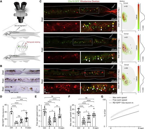
Figure 1. Axon regrowth of propriospinal V2a interneurons is highly correlated with locomotion recovery after SCI (A) Illustration of the adult zebrafish spinal cord injury (SCI) model and rhodamine dextran (RD) retrograde labeling of V2a interneuron with regrown axons in Tg(Chx10:GFP). (B) Spinal cord tissue recovery at 2, 4, and 8 weeks post injury (wpi). (C) Left: images of V2a interneurons with regrown axons (GFP+/RD+, arrowheads) at 2, 4, and 8 wpi. Details are enlarged beneath. Right: two-dimensional kernel density analysis of spatial distribution of V2a interneuron with regrown axons along the dorsoventral and mediolateral aspects of the spinal cord. One dot equals one neuron. N = 3 fish and four segments immediately rostral to the lesion were measured in each fish. (D) Quantification of retrogradely labeled propriospinal V2a interneuron numbers in SCI animals at 2, 4, and 8 wpi, and uninjured fish. One dot equals one fish. (E and F) Free and maximum swimming speed of SCI and uninjured animals. One dot equals one fish. (G) Correlation analysis between the number of V2a interneurons with regrown axons, and free or maximum swimming speeds. Values are presented as a percentage of those measured in uninjured fish. All data are presented as mean ± SD. ?p < 0.05, ??p < 0.01, ???p < 0.001, ????p < 0.0001, significant difference. See also Figure S1 |

