
Fig. 1
- ID
- ZDB-FIG-230204-28
- Publication
- Brown et al., 2021 - A novel gene trap line for visualization and manipulation of erbb3b+ neural crest and glial cells in zebrafish
- Other Figures
- All Figure Page
- Back to All Figure Page
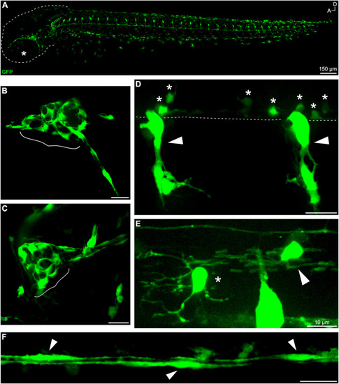
Fig. 1. gSAIzGFFD37A-driven GFP is expressed in neural crest-derived cells and glial subtypes. (A) Whole-mount, in vivo imaging of a gSAIzGFFD37A embryo at 48 hpf (dashed line outlining head; star marking eye). In vivo imaging of fluorescent structures including (B) anterior lateral line ganglion and (C) posterior lateral line ganglion (in brackets) at 48 hpf. Trunk structures include (D) neural crest-derived glia (arrows) and ventral OPCs (stars). Dashed line denotes edge of ventral spinal cord. (E) GFP+ OPC (star) and OL (arrow) at 72 hpf in the spinal cord. (F) GFP+ lateral line Schwann cells at 48 hpf. Scale bars, 20 μm unless otherwise noted. |
Reprinted from Developmental Biology, 482, Brown, E.A., Kawakami, K., Kucenas, S., A novel gene trap line for visualization and manipulation of erbb3b+ neural crest and glial cells in zebrafish, 114-123, Copyright (2021) with permission from Elsevier. Full text @ Dev. Biol.