|
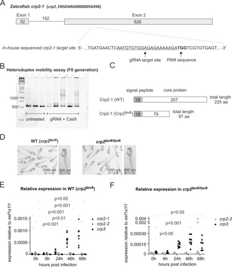
<italic toggle='yes'>crp2-1</italic> knockout zebrafish produce truncated form of Crp2-1 and show compensatory expression of <italic toggle='yes'>crp3</italic>.A) A schematic presentation of the zebrafish crp2-1 (crp2, ENSDARG00000056498) gene structure and the gRNA target site for CRISPR-Cas9 mutagenesis. The target site sequence with depicted gRNA target sequence and protospacer adjacent motif (PAM) site were verified with Sanger sequencing. B) Successful mutagenesis in the embryos injected with target specific gRNA and Cas9 protein was verified with heteroduplex mobility assay. In the heteroduplex mobility assay, multiple bands on polyacrylamide gel after heteroduplex formation indicate the presence of insertion/deletion mutations at the target site. C) Schematic representation of the structure of wild type (WT) Crp2-1 protein and the structure of Crp2tpu6 mutant protein after introduction of +23 nucleotide insertion and premature stop codon by CRIPSR-Cas9 mutagenesis. D) Homozygous crp2tpu6/tpu6 larvae have normal morphology under standard laboratory conditions. The panel shows an overview and a representative image of a single larvae of 2 dpf wild type (WT (crp2tpu6)) and homozygous (crp2tpu6/tpu6) larvae of F3 generation. Images were taken with Lumar V.12 fluorescence stereomicroscope with an exposure time of 100 ms. E) crp2-1 expression is induced in WT (crp2tpu6) larvae at 24 hpi. F) crp3 expression is induced in homozygous crp2tpu6/tpu6 larvae at 24 hpi. In E and F, the expression relative to eef1a1l1 expression was measured with the 2-ΔCt method from the pools of five WT or mutant larvae infected with 241 cfu of S. pneumoniae. The line depicts the median expression level, and the statistical analyses were conducted with Kruskal-Wallis test with Dunn’s multiple comparisons test. Comparisons were only done to 0 h time point of the same gene.
|

