
Figure 4
- ID
- ZDB-FIG-221226-108
- Publication
- Lo et al., 2022 - GTP-Binding Protein 1-Like (GTPBP1l) Regulates Vascular Patterning during Zebrafish Development
- Other Figures
- All Figure Page
- Back to All Figure Page
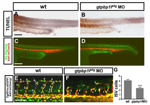
gtpbp1l is required for the proliferation and migration of ISV cells. (A?D) The TUNEL assay and Acridine orange (AO) staining were used to examine apoptotic cells in wild-type (wt) control and gtpbp1l knockdown morphants. (A,B) The number of apoptotic cells (black dots) likely increased in the epidermis of the dorsal region instead of vessel areas in gtpbp1l MO compared to controls at 30 hpf. (C,D) AO staining in Tg (kdrl:mCherry)ci5 fish showed that more apoptotic cells (green dots) were present in the dorsal region of the embryos, but not in vasculature (red fluorescence) after gtpbp1l knockdown. (E,F) At 30 hpf, using transgenic fish Tg(kdrl:mCherryci5; fli1a:negfp y7), the number of ISV endothelial cells can be counted in wt control and gtpbp1l morphants. (G) Quantification of the average numbers of ISV endothelial cells per ISV in wt control (4.1 � 0.8, n = 30 ISVs from 6 embryos) and gtpbp1l morphants (2.5 � 1.3, n = 30 ISVs from 6 embryos). Scale bars are 200 ?m for (A?D) and 100 ?m for (E,F). Data are represented as means � S.D. *** refers to p < 0.0001 by an unpaired Student?s t-test. |
| Fish: | |
|---|---|
| Knockdown Reagent: | |
| Observed In: | |
| Stage: | Prim-15 |