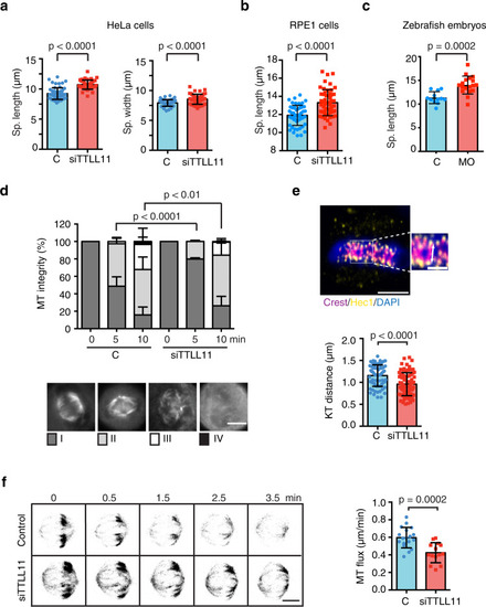
a Quantification of the spindle length and width in control or siTTLL11 HeLa cells. Length: n (control) = 55 cells and n (siTTLL11) = 45 cells. Width: n (control) = 47 cells and n (siTTLL11) = 47 cells. Graphs show one representative experiment of N = 4 independent experiments. Data are presented as mean values +/? SD. b Quantification of the spindle length in control and siTTLL11 hTERT-RPE1 cells. n (control) = 53 cells and n (siTTLL11) = 53 cells. Graphs show one representative experiment of N = 3 independent experiments. Data are presented as mean values +/? SD. c Quantification of the spindle length in control and MO-1 zebrafish embryo dissected cells. n (control) = 28 cells and n (MO) = 26 cells. Graphs show one representative experiment of N = 2 independent experiments. Data are presented as mean values + /? SD. d Quantification of cold-induced K-fibers depolymerization over time in control and siTTLL11 cells. Cells were classified into four categories as shown. n (control[0]) = 100 cells, n (control[5]) = 87 cells, n (control[10]) = 87 cells, n (siTTLL11[0]) = 100 cells, n (siTTLL11[5]) = 105 cells and n (siTTLL11[10]) = 115 cells. P values based on ?2 test with a 95% confidence interval. Scale bar, 15 ?m. e Immunofluorescence image of metaphase plate aligned chromosomes in HeLa cells, showing chromosomes (blue), CREST (magenta, kinetochore), and Hec1 (yellow). In the magnification, the white arrow shows the measured interkinetochore distance. Scale bar, 5 ?m; for zoom, 2 ?m. Quantification of interkinetochore distance. For si-control, n = 72 kinetochore pairs, from 16 cells; for siTTLL11, n = 82 kinetochore pairs, from 18 cells. Scale bar, 5 ?m; in zoom, 2 ?m. f Confocal images of tubulin photoactivated (dark gray) close to the metaphase spindle equator (0 min) over time (min) in control or siTTLL11 cells. The velocity of the poleward flux in control cells (n = 18) and siTTLL11 cells (n = 14). Scale bar, 15 ?m. All graphs correspond to three independent experiments. Data are presented as mean values +/? SD. Unless otherwise indicated P values are based on an unpaired two-sided t test with 95% confidence. Source data are provided as a Source Data file.
|

