
Fig. 1
- ID
- ZDB-FIG-220822-9
- Publication
- DeMarco et al., 2022 - A genetic labeling system to study dendritic spine development in zebrafish models of neurodevelopmental disorders
- Other Figures
- All Figure Page
- Back to All Figure Page
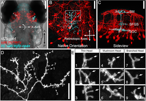
Dendritic spines on PyrN apical dendrites. (A) Dorsal view, whole-brain confocal image volume of an 8 dpf double transgenic Tg(id2b:gal4,uas-e1b:ntr-mCherry) larva injected at the embryo stage with uas:egfp-caax plasmid to generate sparse labeling. Note the single EGFP-labeled PyrN in each tectal lobe. (B) Higher magnification of the maximum projection of neurons labeled in the right tectal lobe of the larva in A. Projection is shown from the dorsal view, with 0° rotation. Note that this view was used for subsequent measurements of dendrite area along the retinotopic axes. (C) Maximum projection of same neuron rotated −50° about the x-axis, to yield an orientation parallel to the tectal layers. Note the clearly stratified neurite morphology with arbors in the SM, SFGS and SGC layers of the tectal neuropil. (D) High magnification view of a subvolume of the SM-targeted dendrite of PyrN in B,C as indicated by the box in B. Note the branched arbor decorated with multiple short protrusions. Numbered arrows indicate spines selected for higher magnification views. (E) 3× magnified views of nine dendritic spines indicated by arrows in D. Note the presence of different types of spine heads: thin, mushroom and branched. Images are representative of 20 PyrNs. Scale bars: 100 µm (A); 25 µm (B,C); 10 µm (D); 1.65 µm (E). |
| Gene: | |
|---|---|
| Fish: | |
| Anatomical Term: | |
| Stage: | Days 7-13 |