FIGURE

Fig 3 s1

ID
ZDB-FIG-220808-27
Publication
Indrischek et al., 2022 - Vision-related convergent gene losses reveal SERPINE3's unknown role in the eye
Other Figures
All Figure Page
Back to All Figure Page
Fig 3 s1

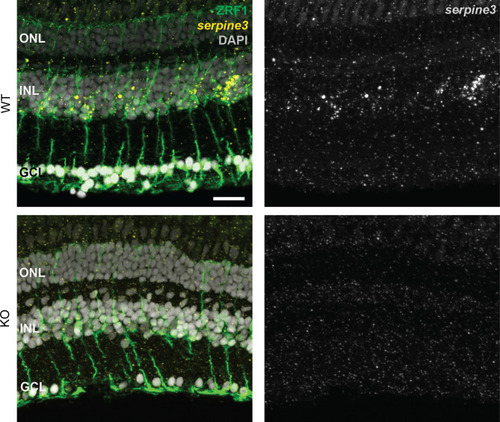
<italic toggle='yes'>Serpine3</italic> expression co-localizes with glial fibrillary acidic protein in the retina of wild type fish.

The fluorescence in situ signal for zebrafish serpine3 is specific for wild type (WT, yellow), where it is in proximity to staining of the ZRF1-antibody (green). This indicates expression of serpine3 by Müller glia cells. Serpine3 signal is not present in homozygous serpine3cbg17 siblings. In the overlay, nuclei (DAPI) are shown in white. ONL – outer nuclear layer, INL – inner nuclear layer, GCL – ganglion cell layer. Scale bar = 20 µm.

Expression Data
Genes:
Antibody:
Fish:
Anatomical Terms:
Stage: Adult

Expression Detail
Antibody Labeling
Phenotype Data
Fish:
Observed In:
Stage: Adult

Phenotype Detail
Acknowledgments
This image is the copyrighted work of the attributed author or publisher, and ZFIN has permission only to display this image to its users. Additional permissions should be obtained from the applicable author or publisher of the image. Full text @ Elife