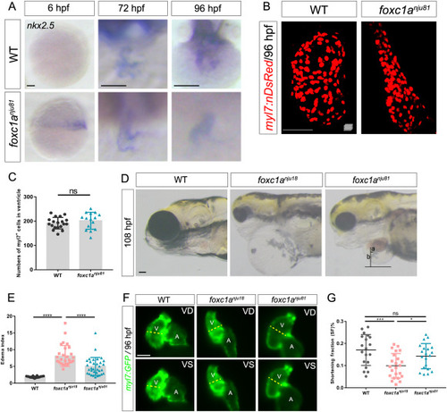
Fig. 5. The defects observed in the foxc1a nju18 ventricle can be partially abated by the conditional knockin of nkx2.5 in foxc1a+ cells. A: The WISH results reveal that the expression of nkx2.5 is rescued in foxc1a nju81 hearts. Sample size: WT (n = 10), foxc1a nju81 (n = 15) at 6 hpf; WT (n = 9), foxc1a nju81 (n = 6) at 72 hpf; WT (n = 14), foxc1a nju81 (n = 7) at 96 hpf. The 6 hpf embryos are lateral views with the animal pole oriented upward. The 72 and 96 hpf embryos are ventral views with the head oriented upward. B: Maximum intensity projections of confocal z-stacks show representative hearts of WT (n = 19) and foxc1a nju81 (n = 15) embryos at 96 hpf. C: A scatter plot shows the numbers of ventricular cells. D: Lateral views of WT, foxc1a nju18, and foxc1a nju81 embryos. a, The distance from the center of the ventricle to the edge of the heart; b, the distance from the center of the ventricle to the edge of the pericardium. E: A scatter plot shows the pericardial edema indexes of the WT (n = 20), foxc1a nju18 (n = 29), and foxc1a nju81 (n = 33) embryos at 108 hpf. F: Representative hearts of foxc1a nju18 and foxc1a nju81 embryos at 96 hpf. G: A scatter plot shows the SF in WT (n = 21), foxc1a nju18 (n = 25), and foxc1a nju81 (n = 21) embryos. ?, P < 0.05; ??, P < 0.01; ???, P < 0.001; ????, P < 0.0001; ns, P > 0.05; Error bars represent the standard deviation. Scale bars, 50 ?m (A, B, F); 100 ?m (D). V, ventricle; A, atrium; VD, ventricular diastole; VS, ventricular systole.
|
