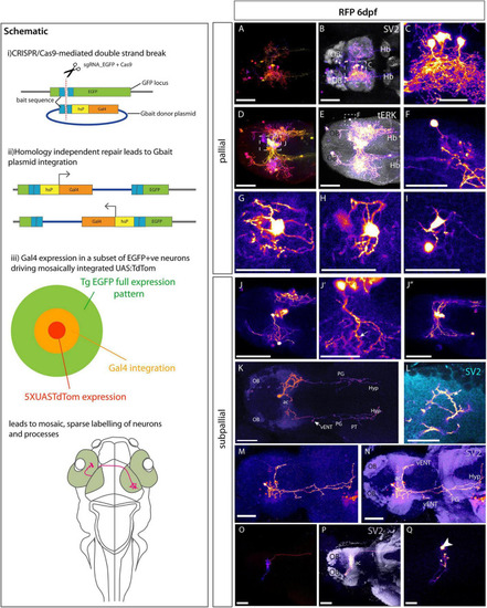
Morphologies of pallial and subpallial neurons. Schematic: Schematic parts (i,ii) adapted from (iii) Shows our adaptation of this method to sparsely label single neurons and their processes by adding 5XUASTdTom to the injection mix. (A?Q) Et(gata2:EGFP)bi105 larvae injected with CRISPR cocktail and labeled with anti-RFP and other markers. (A?I) Pallial Neurons. (A?I) Dorsal views of 6dpf Et(gata2:EGFP)bi105 larvae injected with CRISPR cocktail. Labeled with anti-RFP (FIRE depth color code) (A,D) and FIRE LUT (B,C,E,F), anti-SV2 (gray) (B), anti-tERK (gray) (E). (C) Detail of ?monopolar? dorsal pallial neurons from larva shown in panel (A,B) [area indicated by whisker box in panel (B)]. (F) Detail of single monopolar pallial neuron from larva shown in panels (D,E) [area indicated by whisker box in panel (E)]. (G?I) Three examples of basket shaped cells all found in the dorsal pallium of larva shown in panels (D,E). (J?Q): Subpallial neurons. (J?J?) A cluster of midline subpallial cells from larva shown in panels (D,E) exhibiting elaborate dendritic trees that project throughout the subpallium at the level of the ac. These dendrites exhibit a spiny morphology [arrow in panel (J?)]. (K?N) Ventral view of two different 6dpf, labeled with anti-RFP (FIRE) and anti-SV2 [cyan in panel (L), purple in panel (N)]. (K) Subpallial neuron with cell body just rostral to ac and bilateral projections. Arrow in panel (K) shows branching in the area of the lfb adjacent to the vENT. This cells also extends processes to tuberal and hypothalamic areas. (L) Close-up details of cell processes and dendritic spines (arrow) in the ipsilateral telencephalon from cell shown in panel (K). (M,N) Subpallial neuron also with cell body rostral to ac. It shows elaborated dendrites in the ipsilateral subpallium and one process crossing in the ac to innervate the contralateral telencephalon. Long processes project caudally to likely innervate the ventral entopeduncular nucleus, tuberal area and hypothalamus. (O?Q) A subpallial neuron with a peculiar cocoon morphology [ventral view; FIRE depth color code in panel (O), FIRE LUT in panels (P,Q), anti-SV2 (gray) in panel (P)]. The cell body [arrowhead in panel (Q)] is located within the neuropil area of the ac. The dendrites wrap around the cell within the ac (O,P). A short process innervates the contralateral subpallium rostral to the ac (P). A longer process descends through the fb to contralateral tuberal area (P). Scale bars (A,B,D,E,J?J?,K,M?Q): 50 ?m. Scale bars (C,F?I,L) 25 ?m.

