
Fig. 3 - supplement 1
- ID
- ZDB-FIG-220621-23
- Publication
- Amini et al., 2022 - Amoeboid-like migration ensures correct horizontal cell layer formation in the developing vertebrate retina
- Other Figures
- All Figure Page
- Back to All Figure Page
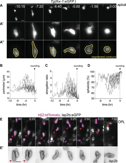
(A) Cytoplasmic deformations of a horizontal cell (HC) migrating from the basal inner nuclear layer (INL) to the HC layer. Cells from Tg(lhx-1:eGFP) transgene embryo were transplanted into wild-type (WT) embryos. White line: apical surface; red dot: tracked HC. Scale bar: 10 ?m. Frames from Video 4. (A?) Close-up of HC in (A). Scale bar: 5 ?m. (A??) Representative sequences of the automated segmented contours of the cell body (yellow line) generated by Icy. (B?D) Cell morphodynamic changes in 12 HCs from seven embryos; (B) perimeter (?m), (C) elongation ratio, (D) sphericity (%). See Figure 3?source data 2. (E-E?) HCs nuclei undergo deformations and exhibit different shapes as they move to their final position. Nuclear envelopes are labeled by lap2b:eGFP (gray), and HCs/PRs are labeled by tr?2:tdTomato (magenta) DNA constructs. Dashed line: outer plexiform layer (OPL); red arrowhead: tracked HC; scale bar: 10 ?m. (E?) Close-up of HC in (E) showing nuclear envelopes (gray). Scale bar: 5 ?m. Time in h:min (A, E). |