
Fig. 1 - supplement 1
- ID
- ZDB-FIG-220621-19
- Publication
- Amini et al., 2022 - Amoeboid-like migration ensures correct horizontal cell layer formation in the developing vertebrate retina
- Other Figures
- All Figure Page
- Back to All Figure Page
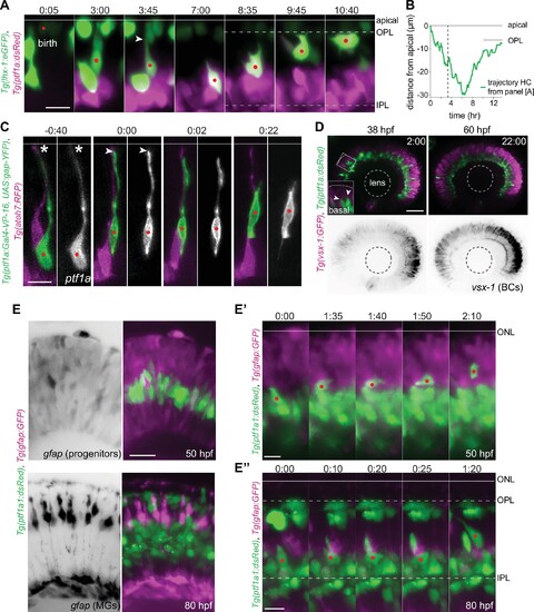
(A) Montage of bidirectional migration of a horizontal cell (HC) from birth to final positioning. Tg(lhx-1:eGFP) labels HCs (green), Tg(Ptf1a:dsRed) marks amacrine cells (ACs) and HCs (magenta). Red dot: tracked HC; arrowhead: HC detachment from the apical surface (line). Scale bar: 10 �m. Frames from Video 1. (B) Trajectory of a migrating HC (depicted in A) from birth to terminal position relative to the apical surface (0 �m). See Figure 1?source data 1. (C) Retraction of HC apical attachment. Tg(ptf1a:Gal4-VP-16, UAS:gap-YFP) labels membrane of HCs (green), and Tg(atoh7:RFP) labels membranes of photoreceptors (PRs) and retinal ganglion cells (RGCs) (magenta). The tracked HC was monitored until it reached the HC layer. Red dot: tracked HC; asterisk: apical attachment; arrowhead: tip of the retracted attachment; line: apical surface. Scale bar: 10 �m. (D) Stills from time-lapse images of a retina before (38 hours post fertilization [hpf]) and after (60 hpf) BC lamination. Tg(ptf1a:dsRed) labels HCs and ACs (green), Tg(vsx1:GFP) marks BCs (magenta). Scale bar: 50 ?m. Higher magnification inset of the outlined region shows two HCs moving apically (arrowheads). (E?E?) HCs do not migrate along radially oriented progenitors or MGs. Tg(Ptf1a:dsRed) marks ACs and HCs (green), Tg(gfap:GFP) labels MGs (magenta). (E) Top: Prior to MG generation (50 hpf). GFAP+ cells are neurogenic progenitors. Bottom: Mature bipolar MG morphology (80 hpf). Scale bar: 20 �m. (E?) Montage of an HC migrating perpendicular to GFAP+ progenitors in 50 hpf. GFAP+ cells are neurogenic progenitors. (E??) Stills of an HC migrating perpendicular to mature MGs in 80 hpf. Scale bar: 10 �m. Line: apical surface; red dot: migrating HC; dotted line: OPL (top), IPL (bottom). Time in h:min (A, C, D, E??E??). |