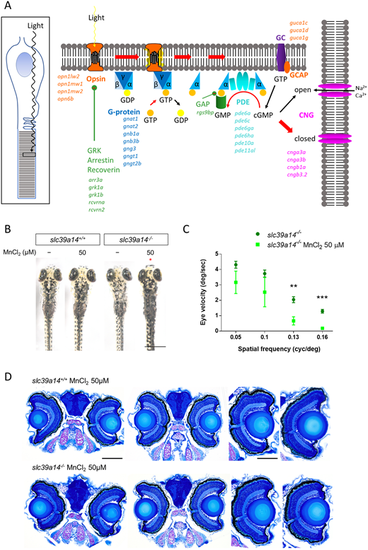
slc39a14?/? mutants develop a visual phenotype upon MnCl2 exposure. (A) Schematic showing the process of phototransduction (Kaupp and Seifert, 2002) with the differentially expressed genes observed in MnCl2-exposed slc39a14?/? mutants indicated in italics. cGMP, cyclic guanosine monophosphate; CNG, cyclic nucleotide-gated non-selective cation channels; GC, guanylyl cyclase; GCAP, guanylate cyclase-activating protein; PDE, phosphodiesterase; GRK, G protein-coupled receptor kinase; GAP, GTPase-activating protein. (B) Dorsal views during light exposure of wild-type siblings (slc39a14+/+, left) and slc39a14?/? larvae (right) at 5?dpf that were unexposed and exposed to 50?�M MnCl2. Note the darker pigmentation of mutants exposed to MnCl2 (red asterisk). Scale bar: 500?�m. (C) Graph showing the eye velocity in response to moving stimuli of different spatial frequencies (average of both eyes) of slc39a14?/? larvae that were unexposed (dark green circles) and exposed to 50?�M MnCl2 (light green squares). Data are presented as mean�s.e.m. from five independent experiments (two-tailed unpaired Student's t-test; **P<0.01; ***P<0.001). (D) Histologic analysis of retinal sections with Richardson?Romeis staining of wild-type siblings (slc39a14+/+, top row) and slc39a14?/? larvae (bottom row) at 5?dpf exposed to 50?�M MnCl2. Scale bars: 200 �m (image of both eyes), 100 �m (image of single eye).
|
