
Fig. 1.
- ID
- ZDB-FIG-220414-16
- Publication
- Takesono et al., 2022 - Estrogens regulate early embryonic development of the olfactory sensory system via estrogen-responsive glia
- Other Figures
- All Figure Page
- Back to All Figure Page
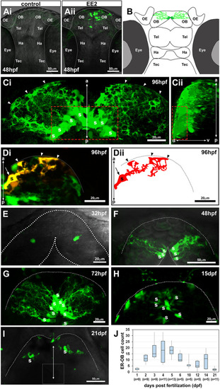
The earliest estrogens/ER-mediated transcriptional activation occurs in a small number of cells in the OB in the zebrafish embryonic brain. (Ai,Aii) Confocal z-projection images of control (Ai) or 17-? ethinylestradiol (EE2, 100 ng/l; Aii)-exposed zebrafish ERE:GFP embryos at 48 hpf. (B) Illustration of the EROB cellular domain in a 4 dpf zebrafish embryo. (Ci,Cii) Confocal z-projection images (Ci, dorsal view; Cii, a 90� rotated view) of EROB cells in EE2-exposed ERE:GFP embryos at 96 hpf. Red dotted rectangles outline the mediodorsal OB; arrowheads show the distal ends of EROB cells. (Di,Dii) Di shows the morphology of a single EROB cell from a confocal z-projection image and Dii illustrates a trace of the cell morphology. The midline is on the left edge; dotted lines show the OB pia; arrowheads show the termini of EROB cells at the OB pia; arrow shows the somata of the EROB cell. (E-I) Confocal z-projection images of EROB cells (green) showing their ontological development in the brain of embryo/larval zebrafish (dorsal view). Each animal stage was exposed to 100 ng EE2/l for up to 4 days before the indicated developmental stage. Dotted lines show the OB pia; white dotted square in I shows the mediodorsal OB. (J) Ontogenic profiles of EROB cell numbers. Boxplot shows median values (middle black bars) and 1st-3rd interquartile ranges (boxes); whiskers extend to the minimum and maximum of the data range within 1.5� the interquartile range. a-p, anterior-posterior axis; d-v, dorso-ventral axis; OB, olfactory bulb; OE, olfactory epithelia; Ha, habenula; S, somata; Tec, tectum; Tel, telencephalon. |