|
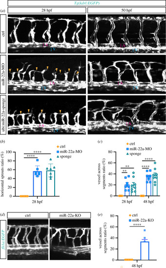
Deficiency of miR-22a caused aberrant vascular networks. (a) Confocal imaging analysis of control MO, miR-22a-MO and miR-22a-sponge-injected Tg(kdrl:EGFP) embryos at 30 hpf and 48 hpf. The arrowheads indicate the aberrant angiogenic sprouts. A: dorsal aorta; V: posterior cardinal vein. (b) Statistics of horizontal sprouts ratio in 30 hpf embryos injected with control MO (n = 7), miR-22-MO (n = 7) and miR-22-sponge (n = 7). One-way ANOVA, **** p < 0.0001. (c) Statistics of embryos with vessel across segments ratio in each group: control MO (n = 6), miR-22a-MO (n = 8) and miR-22-sponge (n = 8). One-way ANOVA, ** p < 0.01; **** p < 0.0001. (d) Confocal imaging analysis of control and miR-22a-KO Tg(fli1a:EGFP-CAAX) embryos at 48 hpf. (e) The statistics of embryos with vessels across segments ratio in each group: control (n = 7) and miR-22-KO (n = 7). t-test, ****, p < 0.0001.
|