FIGURE

Figure 1.

ID
ZDB-FIG-220404-21
Publication
Wiggin et al., 2022 - V3 Interneurons are Active and Recruit Spinal Motor Neurons During In Vivo Fictive Swimming in Larval Zebrafish
Other Figures
All Figure Page
Back to All Figure Page
Figure 1.

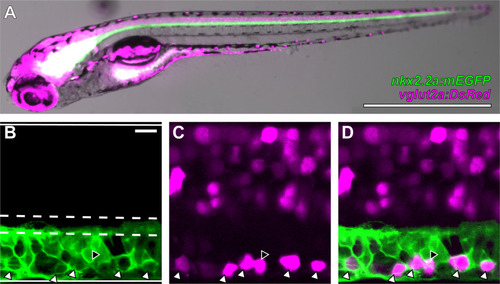
V3-INs are identified by co-localization of vglut2a:DsRed and nkx2.2a:mEGFP transgene expression in neurons located in the ventromedial spinal cord. A, Whole-mount Tg(nkx2.2a:mEGFP)vu17;Tg(vglut2a:DsRed)nns9 double transgenic zebrafish larva. B–D, A representative single confocal optical section of the spinal cord exhibiting: (B) nkx2.2a:mEGFP expression, (C) vglut2a:DsRed expression, and (D) overlayed nkx2.2a:mEGFP and vglut2a:DsRed expression. The boundaries of the spinal cord and central canal are illustrated by solid and dashed white lines, respectively, in panel B. White triangles indicate neurons that co-express both transgenes and the black triangle indicates the only vglut2a:DsRed neuron ventral to the central canal that did not co-express nkx2.2a:mEGFP in the field of view. Scale bars: 1 mm (A) and 10 μm (B–D).

Expression Data

Expression Detail
Antibody Labeling
Phenotype Data

Phenotype Detail
Acknowledgments
This image is the copyrighted work of the attributed author or publisher, and ZFIN has permission only to display this image to its users. Additional permissions should be obtained from the applicable author or publisher of the image. Full text @ eNeuro