
Fig. 9
- ID
- ZDB-FIG-220224-24
- Publication
- Jin et al., 2021 - An animal model for mitochondrial tyrosyl-tRNA synthetase deficiency reveals links between oxidative phosphorylation and retinal function
- Other Figures
- All Figure Page
- Back to All Figure Page
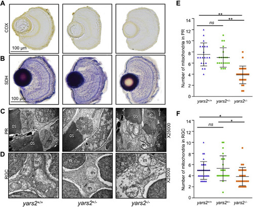
Mitochondrial defects in the zebrafish retina. A and B, assessment of mitochondrial function in the retina by enzyme histochemistry (EHC) staining for COX (A) and SDH (B) in the frozen sections of retina in the yars2+/+, yars2+/?, and yars2?/? larvae at 5 dpf. Scale bar, 100 ?m. C and D, mitochondrial morphology from photoreceptors (PR) (C) and RGC (D) of transmission electron microscopy. Ultrathin sections were visualized with 25,000� magnification. Scale bar, 1 ?m. e, ellipsoid; m, mitochondrion; n, nucleus; os, outer segment of photoreceptors. E and F, quantification of mitochondrial numbers of photoreceptors (E) from yars2+/+ (n = 20), yars2+/? (n = 20), and yars2?/? (n = 25) and RGC (F) from yars2+/+ (n = 32), yars2+/? (n = 24), and yars2?/? (n = 25) zebrafish at 5dpf. Graph details and symbols are explained in the legend to Figure 2. |