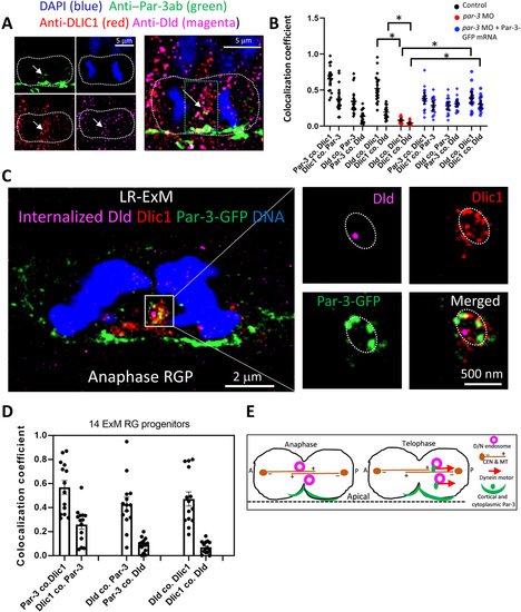
Fig. 5
- ID
- ZDB-FIG-211216-248
- Publication
- Zhao et al., 2021 - Polarized endosome dynamics engage cytoplasmic Par-3 that recruits dynein during asymmetric cell division
- Other Figures
- All Figure Page
- Back to All Figure Page
|
Cytosolic Par-3 together with Dlic1 decorates Dld endosomes.(A) Immunostained anaphase RGP. The green box denotes the area for colocalization analysis (60 × 100 pixels, 0.126-?m pixel size). The white arrows indicate the overlapped cytosolic staining. The MIP of 10 z-planes (0.2-?m z-step) is shown. (B) Quantification of colocalization coefficients (see Materials and Methods) in RGPs. Dld and Dlic1 colocalization is significantly reduced in the par-3?deficient (par-3 MO) RGPs (n = 18, from six embryos of four repeat experiments) compared with control RGPs (n = 20, from five embryos of four repeat experiments). *P < 0.0001 (Dld co. Dlic1, t = 9.56 and df =36; Dlic1 co. Dld, t = 7.27 and df = 36), unpaired two-tailed t test. In the par-3 knockdown RGPs rescued with Par-3-GFP mRNA (par-3 MO + Par-3-GFP mRNA, n = 17, from eight embryos of four repeat experiments), Dld and Dlic1 colocalizations are significantly restored. *P < 0.0001 (Dld co. Dlic1, t = 8.252 and df = 33; Dlic1 co. Dld, t =10.57 and df = 33), unpaired two-tailed t test. Mean with SEM is shown. (C) Left: LR-ExM of anaphase RGP from 28-hpf embryo (par-3-GFP mRNA injected at 16-cell stage and anti?Dld-Atto647 uptake at 26 hpf). Scale bars denote the real biological size. The MIP of 10 z-planes is shown. Right: Enlarged view (MIP of four z-planes) of the endosomal structure (dotted ring-like) containing Dld. Z-step = 0.26 ?m. (D) Statistics of the colocalization coefficient of anti?Par-3, anti-Dlic1, and internalized Dld fluorescence in 14 RGPs (from six embryos of four repeat experiments) processed by LR-ExM. Mean with SEM is shown. (E) Summary model. D/N, delta and Notch; CEN, centrosome; MT, microtubule. |

