Fig. 3
- ID
- ZDB-FIG-211216-246
- Publication
- Zhao et al., 2021 - Polarized endosome dynamics engage cytoplasmic Par-3 that recruits dynein during asymmetric cell division
- Other Figures
- All Figure Page
- Back to All Figure Page
|
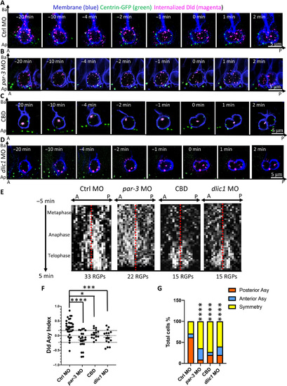
Dld endosome asymmetry is dependent on Par-3 and the dynein motor machinery.(A to D) Time-lapse sequence of images showing Dld endosome dynamics in mitotic RGPs from 28-hpf control MO (A) and embryos deficient for par-3 activity (B), treated with the dynein inhibitor CBD (C), deficient for dynein intermediate light chain 1 (dlic1) (D). Centrosomes are labeled with GFP-centrin, and membrane is labeled with Myr-TdTomato reporter. For (A) to (D), all images shown are the MIP of five confocal z-stacks (1??m z-step). The time interval between each volume of z-stacks is 15 s, and the total acquisition time is 25 min. (E) Kymograph of horizontal projection of (A) to (D) showing distribution of all tracked Dld endosomes along the anteroposterior (A-P) axis (x) over time (y). The red line delineates center point of the axis defined by two centrosomes. (F) Scatter plot showing asymmetry indices in telophase RGPs. Thirty-three control MO RGPs were from 25 embryos of eight repeat experiments, 22 par-3 MO RGPs were from 9 embryos of six repeat experiments, 15 CBD-treated RGPs were from 7 embryos of four repeat experiments, and 15 dlic1 MO RGPs were from 8 embryos of four repeat experiments. The unpaired two tailed t test shows significance between ctrl versus par-3 MO, ****P < 0.0001 (t = 4.706, df = 53); ctrl MO versus CBD, *P = 0.0129 (t = 2.589, df = 46); ctrl versus dlic1 MO **P = 0.0007 (t = 3.645, df = 46). Mean with SEM is shown for each group. (G) Bar graph showing the percentage of RGPs with different patterns of internalized Dld distribution. Disruption of either Par-3 or dynein activity results in a significant decrease of asymmetric posterior Dld segregation. ****P < 0.0001, ?2 test (chi-square = 95.62, df = 6). |

