Figure 1
- ID
- ZDB-FIG-210821-15
- Publication
- Liu et al., 2021 - Insulin-producing β-cells regenerate ectopically from a mesodermal origin under the perturbation of hemato-endothelial specification
- Other Figures
- All Figure Page
- Back to All Figure Page
|
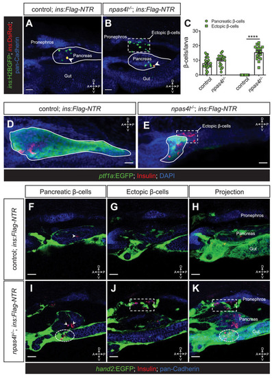
(A, B) Representative confocal projections of the pancreas and neighbouring tissues of control siblings and npas4l?/? Tg(ins:Flag-NTR);Tg(ins:H2BGFP;ins:DsRed) zebrafish larvae at 3 dpf after ?-cell ablation by MTZ at 1?2 dpf, displaying regenerated ?-cells in green and older ?-cells that likely survived the ablation in yellow overlap as the DsRed fluorescence driven by the insulin promoter remained in these cells, at the same time as DsRed had not had enough time to mature in the regenerated ?-cells (arrowheads). The ectopic ?-cells are indicated by white dashed rectangle. Pancreata are outlined by solid white lines. (C) Quantification of the pancreatic or ectopic ?-cells per larva at 3 dpf. ****p<0.0001 (?idák?s multiple comparisons test); n = 24 (control) and 19 (npas4l?/?). Quantification data are represented as the mean ± SEM. (D, E) Representative image projections of the pancreas and neighbouring tissues in control siblings and npas4l-/- Tg(ins:Flag-NTR);Tg(ptf1a:EGFP) larvae at 3 dpf after ?-cell ablation by MTZ at 1?2 dpf, displaying ?-cells in red with immunostaining for insulin and ptf1a:EGFP+ exocrine pancreas in green. Pancreata are outlined by solid white lines. Dashed line outlines ectopic ?-cells in the mesenchyme (E). (F?K) Representative images and projections of the pancreas and neighbouring mesenchyme of control siblings and npas4l-/- Tg(ins:Flag-NTR);Tg(hand2:EGFP) zebrafish larvae at 3 dpf after ?-cell ablation by MTZ at 1?2 dpf, displaying ?-cells in red with immunostaining for insulin and hand2:EGFP+ mesenchyme in green. White arrowheads point to ?-cells in the pancreas (F, I). Dashed rectangles indicate the ectopic ?-cells intermingling with the mesenchyme between the pronephros and the pancreas, without co-expressing insulin and hand2:EGFP (J, K). Selected area in dashed ovals shows other ectopic ?-cells intermingling with the mesenchyme ventral to the pancreas (I, K). Scale bars = 20 ?m. Anatomical axes: D (dorsal), V (ventral), A (anterior), and P (posterior).
|

