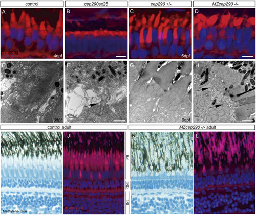
Cep290 morphants and mutants exhibit photoreceptor outer segment length defects. (A-D) BodipyTR (red) staining showed a reduction of photoreceptor outer segment length of 4-dpf cep290ex25 morphants compared with age-matched control morpholino-injected embryos (A,B). MZcep290 mutants also showed a disorganization of photoreceptor outer segment structure at 6 dpf (C,D). (E,F) Electron micrographs of 3-dpf wild-type and cep290ex25 morphant photoreceptors. Although outer segments were found in wild-type retinas at this stage, very few outer segments were observed in cep290ex25 morphants. In some cases, basal bodies and short axonemes could be seen projecting from the cell surface (arrow in F). (G,H) Electron micrographs of 6-dpf heterozygous sibling and MZcep290 mutant photoreceptors showed an abnormal accumulation of cytoplasmic vesicles at the base of the connecting cilium (arrows in H). (I-L) Photoreceptor structure alteration persists until adulthood. Methylene Blue (I,K) and BodipyTR (J,L) staining showed an alteration of photoreceptor outer segment structure and retina lamination in MZcep290−/− adult (1 year old) retinas. Images are representative of >8 embryos/biological replicates. INL, inner nuclear layer; ONL, outer nuclear layer; PR, photoreceptor. Experiments were replicated a minimum of three times. Scale bars: 5 µm (A-D); 50 nm (E-H).
|

