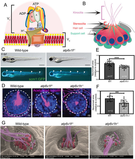
V-ATPase mutant embryos show neuromast defects. (A) Diagram of the V-ATPase holoenzyme complex. The cytosolic V1 domain contains subunits A-H and hydrolyzes ATP, and the Vo domain inserts into membrane and translocates protons (H+). The accessory protein AP1 interacts with the Vo domain. Adapted from Collins and Forgac (2018) and Abbas et al. (2020) under the terms of the CC-BY 4.0 license. (B) Diagram of a lateral view of a zebrafish neuromast with centrally organized hair cells (orange) surrounded by support cells (green). Each hair cell has a single kinocilium (pink), a staircase of stereocilia (red) and a nucleus (blue). (C) Wild-type and atp6v1f−/− embryos at 4 dpf expressing the Tg(scm1:GFP) transgene that labels lateral line neuromasts (white arrowheads). Scale bars: 500 µm. (D) Top-down view of neuromasts in wild-type, atp6v1f−/− and atp6v1h−/− embryos labeled with acetylated tubulin to mark hair cells (magenta) and DAPI to stain nuclei (blue). Dashed line circles indicate the approximate neuromast boundary. (E,F) Quantification of neuromast area in atp6v1f−/− (E) and atp6v1h−/− (F) embryos at 4 dpf relative to wild-type (WT) siblings. n=number of embryos examined. ***P=0.0002, ****P<0.0001 by unpaired Student's t-test with Welch's correction. (G) Pseudo-colored scanning electron micrographs of wild-type, atp6v1f−/− and atp6v1h−/− neuromasts at 4 dpf with pink representing kinocilia and red representing stereocilia. Scale bars: 1 µm.
|
