|
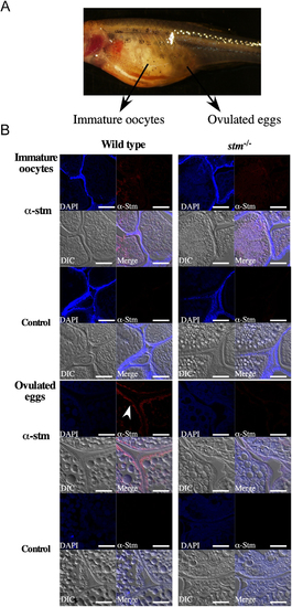
Immunohistochemical observation of Stm. (A) A photograph of stm?/? mutant females possessing ovulated eggs. Frozen sections of immature oocytes and ovulated eggs were prepared from the fish. (B) Immunohistochemical staining results for Stm in WT and in stm?/? mutant immature oocytes (upper panel) and ovulated eggs (lower panel). Frozen sections of the whole bodies of WT and stm?/? mutant zebrafish females were stained with anti-Stm antibodies (?-stm) and DAPI. Differential contrast (DIC) images and merged images of anti-Stm, DAPI and DIC are also indicated (Merge). Control staining (Control) without anti-Stm antibodies of each sample is indicated below. The white arrow indicates signals in anti-Stm staining. The scale bars indicate 50 �m.
|