
FIGURE
Fig. 5
Fig. 5
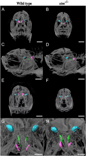
Observation of otoliths by micro CT. Micro CT scan images from anterior (A and B), left lateral (C and D), posterior (E and F) and dorsal (G and H) sides of adult WT zebrafish and the stm ?/? mutant are indicated. Scale bars in A?F are 1 mm. Scale bars in G and H are 500 �m. Three otoliths are indicated in different colours: Lapillus; blue, Sagitta; green, Asteriscus; magenta. Scale bars are 400 �m. |
Expression Data
Expression Detail
Antibody Labeling
Phenotype Data
| Fish: | |
|---|---|
| Observed In: | |
| Stage: | Adult |
Phenotype Detail
Acknowledgments
This image is the copyrighted work of the attributed author or publisher, and
ZFIN has permission only to display this image to its users.
Additional permissions should be obtained from the applicable author or publisher of the image.
Full text @ RAF