|
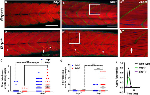
<italic>fkrp</italic><sup><italic>−/−</italic></sup> fish possess a basement membrane failure phenotype.a–d Muscle fibre-crossing and detachment phenotypes at the muscle basement membrane of fkrp−/− mutant larvae. a–b” Z-projected images from the entire mediolateral extent of the myotome centred on the region of the anal pore. Phalloidin conjugated to TRITC (red) stains for f-actin as a marker of skeletal muscle and paxillin (green) visualises vertical myosepta muscle basement membrane integrity. White arrowheads: muscle fibres crossing at defective muscle basement membranes, asterisks: muscle fibre detachment. 3 dpf, scale bar = 100 μm. 5 dpf, scale bar = 75 μm, white boxes in a’, b’ denote the high-magnification views presented in a” and b”. c, d Quantitation of fibre detachment (c) and basement membrane crossing events (d) per myosepta in wild-type (n = 9) or fkrp mutant larvae (n = 9) over five independent experiments, analysed using two-way ANOVA multiple comparisons assuming non-parametric data, the significance of ***(P = < 0.0001), error bars represent SEM. e Physiological analysis of muscle function. The maximum active force (mN) generated over a specific time interval (ms) was measured in individual genotypes of 6 dpf larvae represented in single-twitch recordings of fkrp+/+ sibling (sib) black line (n = 8), fkrp−/− green line (n = 6) and dag1−/− purple line (n = 5).
|