
Fig. 4
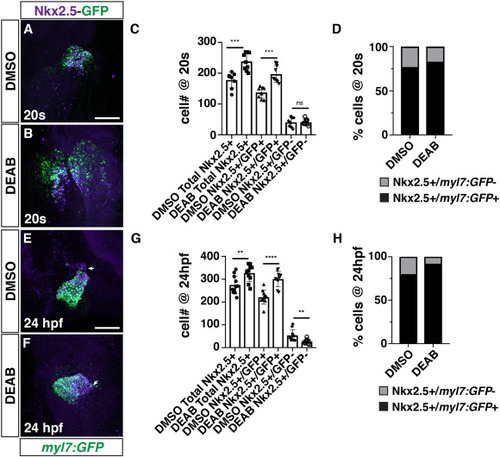
RA restricts the size of the FHF and maintains the SHF progenitor population in DEAB-treated embryos. (A, B) IHC for Nkx2.5 (purple) and GFP (myl7:GFP - green) in DMSO- and DEAB-treated embryos at the 20s stage (19 hpf). The cardiac cone was sometimes delayed in forming in DEAB-treated embryos relative to DMSO-treated control embryos. Views are dorsal with anterior up. Scale bar ? 100 ??m. (C) Quantification of the number of Nkx2.5+/GFP+ and Nkx2.5+/GFP- cells in DMSO- and DEAB-treated embryos at the 20s stage (19 hpf). ??? indicates p ?? ?0.0004. (D) Percentage of Nkx2.5+/GFP+ and Nkx2.5+/GFP- cells within the hearts of DMSO- and DEAB-treated embryos at the 20s stage (19 hpf). (E, F) IHC for Nkx2.5 (purple) and GFP (myl7:GFP - green) in DMSO- and DEAB-treated embryos at 24 hpf. Frontal views with the arterial pole up. Arrows indicate the border between Nkx2.5+/GFP+ and Nkx2.5+/GFP- cells. Scale bar ? 100 ??m. (G) Quantification of the number of Nkx2.5+/GFP+ and Nkx2.5+/GFP- cells in DMSO- and DEAB-treated embryos at 24 hpf. ?? indicates p ?? ?0.0060, ???? indicates p ?< ?0.0001. (H) Percentage of Nkx2.5+/GFP+ and Nkx2.5+/GFP- cells within the hearts of DMSO- and DEAB-treated embryos at 24 hpf. |
Reprinted from Developmental Biology, 473, Duong, T.B., Holowiecki, A., Waxman, J.S., Retinoic acid signaling restricts the size of the first heart field within the anterior lateral plate mesoderm, 119-129, Copyright (2021) with permission from Elsevier. Full text @ Dev. Biol.