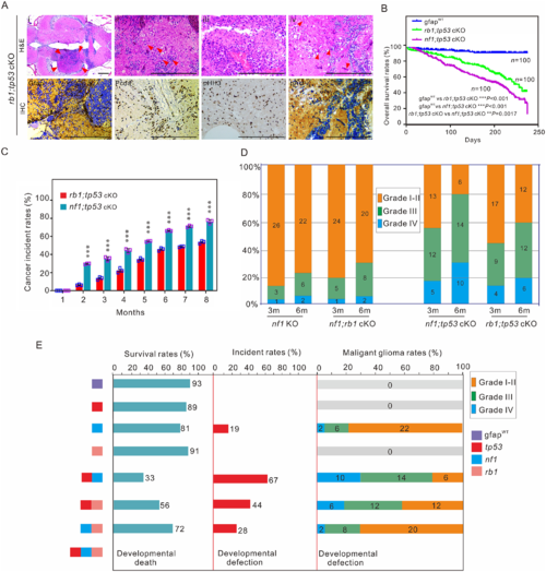
Tp53 mutation is critical for poor prognosis in zebrafish. (A) Histological examination of gliomas in 3-month-old rb1;tp53 cKO fish. (i?iv) Haematoxylin and eosin (H&E) staining showed the generated tumours (arrowheads; i), multinucleated giant cells (arrowheads; ii), the typical gliomatosis phenotype (iii), and vascularity (arrowheads; iv). Immunohistochemistry staining was performed to examine tumour-relevant indicators, including Gfap, Pcna, pHH3, and pAkt, in brain tissues of rb1;tp53 cKO fish. Scale bars = 100 ?m. (B and C) Overall survival rates and cancer incidences of rb1;tp53 cKO and nf1;tp53 cKO fish, respectively (n?=?100 for each group). The glioma formation was preliminarily estimated based on the ?bending-body? phenotype, and confirmed by haematoxylin and eosin staining. (D) Malignancy of the tumours derived from the fish with various mutations at 3 or 6 months of age (n?=?30 for each group). (E) Summary of the survival rates, cancer incidences, and malignancy of the generated gliomas in fish lines with various mutations at 6 months of age. Data shown as mean � SEM. ***P?
|
