|
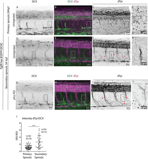
Microtubules of secondary sprouts are selectively detyrosinated. (A-I?) Immunostainings using antibody detecting the glutamate amino acid of detyrosinated microtubules (dTyr) during primary (A-C) and secondary (D-I) sprouting in uninjected (A-F) and plc? KD (G-I) Tg[fli1ep:EGFP-DCX] embryos, labelling all endothelial microtubules (DCX). C?,F? and I? are magnifications from boxed region in C,F,I, respectively. plc? KD embryos (G-I?) show reduced primary sprouting, facilitating the visualization and quantification of dTyr signal specifically in secondary sprouts. Arrowheads indicate secondary sprouts. (J) Quantification of the dTyr/DCX signal intensities in primary sprouts of control MO-injected embryos, and secondary sprouts from plc? MO-injected embryos. n=52 control primary sprouts from 18 embryos, n=24 plc? KD secondary sprouts from 12 embryos, from one experiment. ****<0.00001 (Mann?Whitney test). Pictures are representative of 3 replicated experiments.
|