|
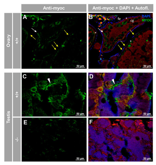
Immunohistochemical detection of myoc in the reproductive system of adult zebrafish. Tissue sections (14 μm) of adult (7 months) wild-type ovary (A,B) and testis (C,D) and KO myoc testis (E,F) were incubated with an anti-myocilin (TNT) primary antibody, followed by Cy2-conjugate goat anti-chicken IgY secondary antibody (green signals). Note that ovaries from −/− animals were not available because of the absence of females with this genotype. White and yellow arrows indicate cortical granules and follicular epithelium-associated immunoreactivity, respectively. White arrowheads show immunolabeling in the seminiferous epithelium (C,D). Red signals correspond to tissue autofluorescence. The images are representative of the result observed in three tissue sections from three animals. +/+: wild-type; −/−: myoc KO. cg: cortical granules. fe: follicular epithelium. *: zona radiata. Negative controls are shown in Figure S6.
|