
Figure 7—figure supplement 1.
- ID
- ZDB-FIG-210220-39
- Publication
- Fontenas et al., 2021 - Spinal cord precursors utilize neural crest cell mechanisms to generate hybrid peripheral myelinating glia
- Other Figures
-
- Figure 1
- Figure 1—figure supplement 1—source data 1.
- Figure 1—figure supplement 2—source data 1.
- Figure 1—figure supplement 3.
- Figure 2—figure supplement 1.
- Figure 2—figure supplement 1.
- Figure 3—figure supplement 1.
- Figure 3—figure supplement 1.
- Figure 3—figure supplement 2.
- Figure 4
- Figure 5—figure supplement 1—source data 1.
- Figure 5—figure supplement 1—source data 1.
- Figure 6—figure supplement 1.
- Figure 6—figure supplement 1.
- Figure 6—figure supplement 2.
- Figure 7—figure supplement 1.
- Figure 7—figure supplement 1.
- Figure 8—figure supplement 1.
- Figure 8—figure supplement 1.
- Figure 8—figure supplement 2—source data 1.
- All Figure Page
- Back to All Figure Page
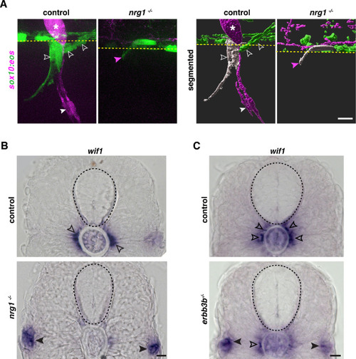
(A) Motor exit point of sox10:eos WT and nrg1 -/- siblings photoconverted at 48 hpf and imaged at 3 dpf showing the presence of MEP glia (outlined arrowheads) and Schwann cells (SCs) (arrowhead) in a control larva and the presence of an oligodendrocyte membrane extension (pink arrowhead) in a nrg1 -/- larva that lacks MEP glia and SCs. Yellow dashed lines denote the edge of the spinal cord. (B) In situ hybridization showing wif1+ MEP glia along motor nerve root axons in a WT control larva (outlined arrowheads, n = 24 larvae) and the absence of wif1+ MEP glia along motor nerve root axons in a nrg1 mutant larva at 3 dpf (n = 16 larvae). Arrowheads indicate the presence of wif1+ cells near the lateral line nerve in a nrg1 mutant larva at 3 dpf. (C) In situ hybridization showing wif1+ MEP glia along motor nerve root axons in a WT control larva (outlined arrowheads, n = 20 larvae) and the absence of wif1+ MEP glia along motor nerve root axons in an erbb3b mutant larva at 3 dpf (n = 14 larvae). Arrowheads indicate the presence of wif1+ cells near the lateral line nerve in an erbb3b mutant larva at 3 dpf. Scale bar (A–C), 10 μm. |