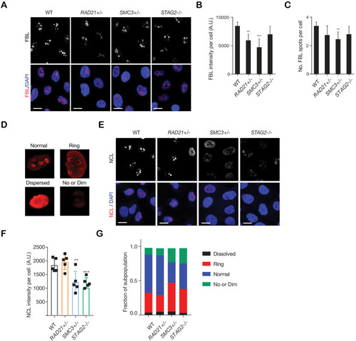
Figure 3—figure supplement 1.
- ID
- ZDB-FIG-201223-26
- Publication
- Chin et al., 2020 - Cohesin mutations are synthetic lethal with stimulation of WNT signaling
- Other Figures
-
- Figure 1.
- Figure 2
- Figure 2—figure supplement 1.
- Figure 2—figure supplement 2.
- Figure 3
- Figure 3—figure supplement 1.
- Figure 3—figure supplement 2.
- Figure 3—figure supplement 3.
- Figure 4
- Figure 4—figure supplement 1.
- Figure 4—figure supplement 2.
- Figure 5
- Figure 5—figure supplement 1.
- Figure 5—figure supplement 2.
- Figure 5—figure supplement 3.
- Figure 6
- Figure 6—figure supplement 1.
- Figure 6—figure supplement 2.
- Figure 7.
- All Figure Page
- Back to All Figure Page
|
Cohesin-deficient cells show altered nucleolar morphology. ( |

