
Figure 2
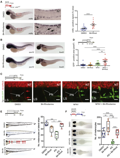
Inhibition of Prl3 Leads to Premature Melanoblast Expansion during Regeneration (A) (B) (C) Confocal imaging of the MSC lineage during treatments specified. (D) Quantification of area of GFP expression (pixels2) on individual peripheral nerves of 84 hpf (E) Lateral stripe melanocytes following specified treatments. AG.: AG1478. B4-Rhod: B4-Rhodanine. Red arrows: missing melanocytes; faint melanocytes from opposite side of the body are also visible. Quantification of melanocytes on the lateral stripe of embryos at 96 hpf, ∗p = 0.0364; ∗∗∗p = 0.0003 and 0.0006 for DMSO versus AG. and AG. versus AG. + B4-Rhod, respectively, ANOVA using Tukey’s analysis. (F) Melanocyte regeneration in |
Reprinted from Developmental Cell, 54(3), Johansson, J.A., Marie, K.L., Lu, Y., Brombin, A., Santoriello, C., Zeng, Z., Zich, J., Gautier, P., von Kriegsheim, A., Brunsdon, H., Wheeler, A.P., Dreger, M., Houston, D.R., Dooley, C.M., Sims, A.H., Busch-Nentwich, E.M., Zon, L.I., Illingworth, R.S., Patton, E.E., PRL3-DDX21 Transcriptional Control of Endolysosomal Genes Restricts Melanocyte Stem Cell Differentiation, 317-332.e9, Copyright (2020) with permission from Elsevier. Full text @ Dev. Cell