Fig. S2
|
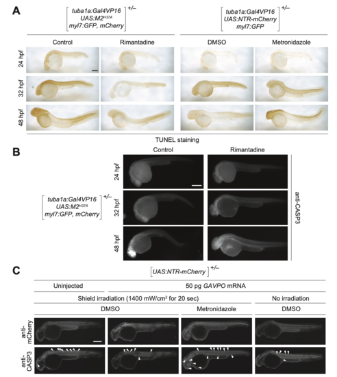
M2H37A- and NTR-induced cell death in zebrafish embryos. (A) Heterozygous Tg(tuba1a:Gal4VP16; UAS:M2H37A;myl7:GFP,mCherry) and Tg(tuba1a:Gal4VP16;UAS:NTR-mCherry;myl7:GFP) embryos were cultured in medium supplemented with DMSO, 100 μg/mL rimantadine, or 5 mM metronidazole at 10 hpf. At 48 hpf, the embryos were fixed, and apoptotic cells visualized by TUNEL staining. TUNEL-positive cells were limited to the CNS at 48 hpf in NTR-expressing embryos treated with metronidazole, but they were observed outside the CNS in M2H37A-express- ing embryos cultured without rimantadine. (B) Heterozygous Tg(tuba1a:Gal4VP16; UAS:M2H37A;myl7:GFP,mCherry) embryos cultured in the presence or absence of 100 μg/mL rimatidine, fixed, and immunostained for activaterd caspase-3 (CASP3). (C) Heterozygous Tg(UAS:NTR-mCherry) zygotes were injected with the designated amount of GAVPO mRNA, and their embryonic shields were irradiated at 6 hpf. The embryos were then cultured in the absence or presence of 5 mM metronidazole from 10 to 32 hpf, fixed, and immunostained for mCherry and activated CASP3 (arrowheads). GAVPO-, light-, and metronidazole-dependent caspase-3 activation was observed. Embryo orienta- tions: lateral view, anterior left. Scale bars: 300 μm. |

