
Fig. s5
- ID
- ZDB-FIG-200611-34
- Publication
- Santos-Ledo et al., 2020 - Alternative splicing of jnk1a in zebrafish determines first heart field ventricular cardiomyocyte numbers through modulation of hand2 expression
- Other Figures
- All Figure Page
- Back to All Figure Page
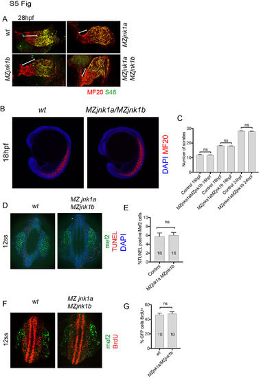
Ventricular hypoplasia in MZjnk1a/MZjnk1b mutants. (A) Cardiac morphology at 28 hpf. All ventricular cardiomyocytes are labelled by MF20 antibody (red) and atrial cells by both MF20 and S46 antibody (appear yellow). Normal appearances seen in wildtype (wt) embryos. Ventricular hypoplasia (indicated by size of white bar) is seen in MZjnk1a null mutants but is not seen in MZjnk1b null mutants. There is no additional ventricular hypoplasia in MZjnk1a/MZjnk1b double mutants. (B) MF20 antibody staining to identify somites/skeletal muscle blocks in wild type (wt) and MZjnk1a/MZjnk1b mutants (C) Somite counting excludes global somatic developmental delay in MZjnk1a/MZjnk1b null mutants. (D,E) TUNEL labelling at 12ss stage in wild type (wt) and MZjnk1a/MZjnk1b mutants. Myocytes in the ALPM are identified by Mef2 antibody. The percentage TUNEL positive cells in Mef2 population is unchanged in MZjnk1a/MZjnk1b null mutants. (F,G) BrdU incorporation in cardiomyocytes from 10ss to 12ss. Quantification shows that there is no difference in BrdU incorporation index in MZjnk1a/MZjnk1b null mutants compared to wild type (wt) controls. ns = nonsignificant. |