
Fig. 6
- ID
- ZDB-FIG-200601-9
- Publication
- Lombardo et al., 2019 - Morphogenetic control of zebrafish cardiac looping by Bmp signaling
- Other Figures
- All Figure Page
- Back to All Figure Page
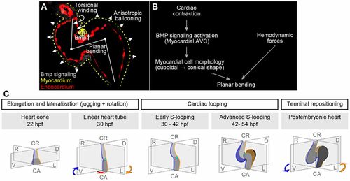
Model of zebrafish cardiac looping. (A) Summary of cellular processes that contribute to zebrafish S-looping morphogenesis: (1) BMP-dependent planar bending (white line at heart midline) of the heart tube due to cuboidal-to-conical cell shape changes of superior AVC cardiomyocytes (yellow triangles); (2) myocardial anisotropic ballooning due to region-specific cell morphology changes causes an expansion of the two cardiac chambers (white arrowheads); and (3) ventricular rightward-torsional winding (twisted arrows at ventricular outflow tract) is most pronounced at the cranial pole of the heart. Bmp signaling (false-colored gray) with strong activation at myocardial superior AVC, myocardium (yellow dotted lines) and endocardium (false-colored red). (B) Summary of cell-intrinsic and -extrinsic factors involved in cardiac planar bending of the heart tube. Cardiac contractility is essential for a strong Bmp signaling pathway activation within myocardial cells at the superior AVC region. This Bmp activation induces myocardial changes from cubical to conical cell shapes and results in an S-planar bending of the heart tube. In addition, hemodynamic forces (shear stress and retrograde flow fraction) affect Bmp signaling-independent planar bending. (C) During zebrafish cardiac looping morphogenesis, two separate processes occur in parallel: (1) planar S-looping along the mid-sagittal body axis and (2) lateral looping during which the ventricle is positioned towards the right and the atrium towards the left (D-looping). The mid-sagittal plane is in grey; original right and left sides of the heart are marked in blue and yellow, respectively. Under some experimental conditions, inverted L-looping can also occur. In comparison with lung-breathing vertebrates, the zebrafish atrium turns around the cardiac cranio-caudal axis. The rotation of the atrium turns the cross-section plane of the AVC along the cranio-caudal axis towards the right. This movement is ‘corrected’ during repositioning, which places the ventricle and atrium into an almost bilateral symmetrical position again. |