
Fig. 5
- ID
- ZDB-FIG-200601-8
- Publication
- Lombardo et al., 2019 - Morphogenetic control of zebrafish cardiac looping by Bmp signaling
- Other Figures
- All Figure Page
- Back to All Figure Page
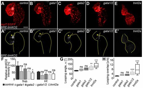
Cardiac contractility is required for Bmp signaling at the superior AVC. (A-E) Reconstructions of confocal z-stacks of hearts at 54 hpf in (A) wild-type, (B) gata1 morphant, (C) gata2 morphant, (D) gata1/2 double morphant or (E) tnnt2a morphant embryos. Myocardial tissue is marked by expression from the Tg(mly7:EGFP)twu34 reporter (false-colored red) and Bmp reporter Tg(BRE:dmKO2)mw40 (false-colored gray). (A′-E′) Bmp reporter Tg(BRE:dmKO2)mw40 expression. The outline of the heart is delineated by dotted lines. Asymmetric Bmp signaling activity within the superior AVC is marked by yellow asterisks. (F-H) Quantifications of (F) relative dmKO2 intensities at the superior (Sup.) and inferior (Inf.) AVC (data in Table S5), (G) looping angle (α) (data in Table S1), and (H) index a/b (data in Table S1) were measured in gata1, gata2, gata1/2 and tnnt2a morphant versus wild-type hearts. Under abnormal hemodynamic conditions (gata1, gata2 and gata1/2 morphant hearts) the relative dmKO2 intensity is not significantly affected. However, asymmetric Bmp signaling activity is lost in the absence of cardiac contractility. The looping angle (α) (G) and the index a/b (H) are significantly increased in gata1/2 and tnnt2a morphant versus control hearts (statistical analysis in Table S1). In G,H, the limits of the boxes indicate the range between the first quartile (25th percentile) and the third quartile (75th percentile). The line inside box indicate the median value. The error bars indicate the maximum and minimum values. Data are mean±s.d. in F; ns, not significant; *P≤0.05; **P≤0.01; ***P≤0.001. Scale bar: 50 μm. |