
Figure 2.
- ID
- ZDB-FIG-200530-30
- Publication
- Ye et al., 2020 - Yap-lin28a axis targets let7-Wnt pathway to restore progenitors for initiating regeneration
- Other Figures
-
- Figure 1
- Figure 1—figure supplement 1.
- Figure 1—figure supplement 2.
- Figure 2—figure supplement 1.
- Figure 2—figure supplement 2.
- Figure 2.
- Figure 3—figure supplement 1.
- Figure 3—figure supplement 2.
- Figure 3.
- Figure 4
- Figure 4—figure supplement 1.
- Figure 5
- Figure 5—figure supplement 1.
- Figure 6
- Figure 6—figure supplement 1.
- Figure 7.
- All Figure Page
- Back to All Figure Page
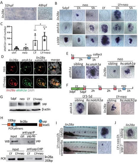
(A) Lin28a was not expressed in the developing lateral line primordium or neuromast. (B) Treatment with Notch inhibitor LY411575 from 3dpf to 5dpf increased expression atoh1a and s100t. More lin28a expression was observed in LY+neo-1h compared with neo-1h. No lin28a was detected at 5 hr post LY+neo or neo. (C) The number of atoh1a-transcribed cells detected by in situ were higher at 1 hr post LY+neo compared with LY. (D) Double fluorescent in situ showed that lin28a was co-expressed with atoh1a post neo or LY+neo. (E) Induction of lin28a post injury was increased when atoh1a was overexpressed in hs:atoh1a. (F) Lin28a expression was completely blocked when atoh1a was inhibited in hs:notch1a. (G) Western blot results showed that LY+neo induced higher yap expression compared with neo alone. (H) Motifs of yap and tead1 (co-transcriptional factor of yap) binding sites were predicted near lin28a transcriptional start site. CHIP-PCR results verified that yap directly binds the predicted motif. (I and J) Inhibition of yap using verteporfin or yap mutant blocked lin28a induction post LY+neo. Scale bar equals 10 ?m. |