
Fig. 7
- ID
- ZDB-FIG-200131-23
- Publication
- Fukuda et al., 2019 - Mechanical Forces Regulate Cardiomyocyte Myofilament Maturation via the VCL-SSH1-CFL Axis
- Other Figures
- All Figure Page
- Back to All Figure Page
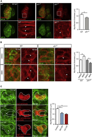
cfl1 Is Essential for Cardiomyocyte Myofilament Maturation (A) MF20 immunostaining and phalloidin staining of 82 hpf WT and cfl1?/? hearts. cfl1?/? CMs exhibit disorganized myofilaments than WT (arrowheads). Myofilament thickness measured in 82 hpf ventricles (n = 4 ventricles). (B) MF20 immunostaining and phalloidin staining of 60 hpf WT and cfl1?/? hearts treated with DMSO or IBMX from 36 to 60 hpf. IBMX treatment leads to myofilament disruption in cfl1?/? CMs than WT (arrowheads). Myofilament thickness measured in 60 hpf ventricles (n = 4 ventricles). (C) 3D images of 100 hpf ventricles. Overexpression of either DN Cfl1 or CA Cfl1 leads to myofilament disruption (arrowheads) compared to control. Myofilament thickness measured in 100 hpf ventricles (n = 6 CMs). Error bars, SEM. ?p < 0.05 by ANOVA followed by Tukey?s HSD test. Scale bars, 20 ?m. |
Reprinted from Developmental Cell, 51(1), Fukuda, R., Gunawan, F., Ramadass, R., Beisaw, A., Konzer, A., Mullapudi, S.T., Gentile, A., Maischein, H.M., Graumann, J., Stainier, D.Y.R., Mechanical Forces Regulate Cardiomyocyte Myofilament Maturation via the VCL-SSH1-CFL Axis, 62-77.e5, Copyright (2019) with permission from Elsevier. Full text @ Dev. Cell