
Fig. 2
- ID
- ZDB-FIG-191118-21
- Publication
- Große et al., 2019 - Zebrafish Wtx is a negative regulator of Wnt signaling but is dispensable for embryonic development and organ homeostasis
- Other Figures
- All Figure Page
- Back to All Figure Page
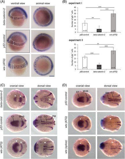
Wtx inhibits the canonical Wnt pathway in early zebrafish development. Whole mount in situ hybridization to detect mRNA expression changes indicative of modulated Wnt/??catenin signaling. The wtx depletion phenotypes were compared with p53?control MO?injected embryos and beta?catenin?2 MO?injected embryos. A, Examination of gfp expression in zebrafish embryos heterozygous for a ??catenin responsive transgene Tg(TOP:dGFP) at shield stage (6 hpf). Embryos are shown from a ventral view with the animal pole at top and from an animal view with dorsal to the right. Depletion of ??catenin2 resulted in reduced transgene expression whereas embryos injected with the wtx MO displayed increased number of ??catenin responsive cells in an expanded area within the ventrolateral margin. GFP?positive cells are recognizable as blue spots inside the areas which are marked with dashed lines in the ventral views; arrows illustrate the expansion of the transgene expressing area in wtx morphants. The diffuse staining around the edge of the embryo is not specific. B, Quantification of the data shown in (A). Images were blinded and the numbers of gfp positive cells were counted. Results from two independent experiments are shown. Error bars represent SD. P values according to the two?tailed t?test after Bonferroni correction: ** P?<?.01, *** P?<?.001. C and D, Expression analysis of markers for lateral mesoderm and neuroectoderm by using a pax2a/myod1/six3b probe mixture on 12 hpf zebrafish embryos. Embryos are shown in a cranial and dorsal view with anterior to the left. C, Knockdown of ??catenin2 caused a lateral shortening of the pax2a expression domain in the MHB and reduced expression of six3b in the optic primordia (white arrows). The myod1 domain in the somites is fused (white asterisk) and absent in the somitic and presomitic mesoderm. In contrast wtx morphants show expanded expression of pax2a, myod1 and six3b(white arrowheads). For ?beta?catenin?2, ?p53 control? and ?wtx (ATG)? 6, 3 and 11 embryos were analyzed, respectively. (D) Wtx (ATG) and wtx (splice) morphants show expanded expression of six3b (white arrows) and myod1 (white arrowheads). For ?p53 control?, ?wtx (ATG)? and ?wtx (splice)? 7, 13 and 9 embryos were analyzed, respectively. EF: eye field; MHB: midbrain?hindbrain boundary; PM: presomitic mesoderm; SM: somitic mesoderm; SO: somites. Scale bar represents 150??m |
| Genes: | |
|---|---|
| Fish: | |
| Knockdown Reagents: | |
| Anatomical Terms: | |
| Stage Range: | Shield to 5-9 somites |
| Fish: | |
|---|---|
| Knockdown Reagents: | |
| Observed In: | |
| Stage Range: | Shield to 5-9 somites |