Fig. 7
|
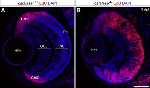
Catalase Endogenous Expression Was Also Required for the Switch of RPC from Proliferation to Differentiation during Normal Retinal Development (A and B) Catalase null-mutant embryos have an increased number of proliferative cells in comparison to their wild-type siblings as observed by EdU incorporation from 2 to 3 days postfertilization (dpf). This increase was particularly striking in the region of the central retina, where by 2 dpf post-mitotic neurons are located in normal condition (GCL, ganglion cell layer; INL, inner nuclear layer; Ph, photoreceptor cell layer; and CMZ, ciliary marginal zone). Scale bars, 35 μm in (A) and (B). |
| Fish: | |
|---|---|
| Observed In: | |
| Stage: | Protruding-mouth |
Reprinted from Developmental Cell, 50(1), Albadri, S., Naso, F., Thauvin, M., Gauron, C., Parolin, C., Duroure, K., Vougny, J., Fiori, J., Boga, C., Vriz, S., Calonghi, N., Del Bene, F., Redox Signaling via Lipid Peroxidation Regulates Retinal Progenitor Cell Differentiation, 73-89.e6, Copyright (2019) with permission from Elsevier. Full text @ Dev. Cell

