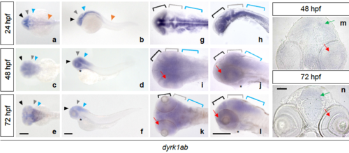
Fig. S3
|
dyrk1ab is expressed in the developing brain region. By WISH (whole mount in situ hybridization), dyrk1ab was expressed in the forebrain (black arrowheads, a-f; black brackets, g-l), the midbrain (gray arrowheads, a-f; gray brackets, g-l), the hindbrain (blue arrowheads, a-f; blue brackets, g-l) at 24, 48 and 72 hpf and the spinal cord (orange arrowheads, a and b) at 24 hpf. It was also detected in the heart (asterisks, d, f, j and l) and in the retina (red arrows, i-l) at 48 and 72 hpf. (m and n) Sectioned images of WISH embryos showed the expression of dyrk1ab in the tectum (green arrows) and the retina (red arrows) at 48 hpf and 72 hpf. Scale bars: 200 μm in (a-l) and 50 μm in (m and n) |
| Gene: | |
|---|---|
| Fish: | |
| Anatomical Terms: | |
| Stage Range: | Prim-5 to Protruding-mouth |

