
Fig. 3
- ID
- ZDB-FIG-190801-49
- Publication
- Jiménez-Amilburu et al., 2019 - The transmembrane protein Crb2a regulates cardiomyocyte apicobasal polarity and adhesion in zebrafish
- Other Figures
- All Figure Page
- Back to All Figure Page
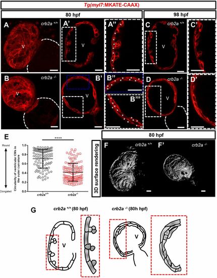
crb2a−/− hearts display disrupted compact wall integrity and fail to form trabeculae. (A-B″) Confocal images (maximum intensity projections) of 80?hpf Tg(myl7:MKATE-CAAX) crb2a+/+ (A-A″) and crb2a−/− (B-B″) hearts. (A-D′) Confocal images (mid-sagittal sections) of Tg(myl7:MKATE-CAAX) hearts of crb2a+/+ (A-A″, n=8; C,C′, n=8) and crb2a−/− (B-B″, n=14; D,D′, n=14) larvae at 80 and 98?hpf. Asterisks in A″,B″ and B? indicate individual CMs in the compact wall. (E) Circularity of ventricular CMs (outer curvature) in 80?hpf crb2a+/+ (n=7 hearts) and crb2a−/− (n=10 hearts) larvae. Each point represents data from an individual CM. Data are mean±s.e.m. ****P<0.0001 by Student's t-test. (F,F′) 3D surface rendering images of 80?hpf crb2a+/+ (F) and crb2a−/− (F′) ventricles. (G) Schematic illustration of the myocardial wall in 80?hpf crb2a+/+ and crb2a−/− ventricles. V, ventricle. |