|
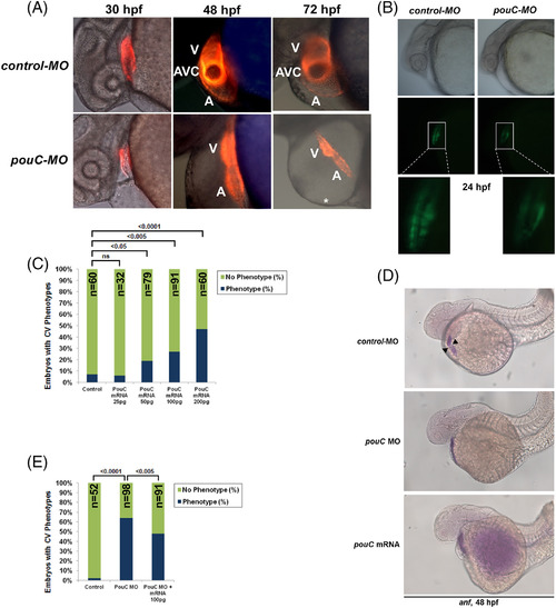
Precise levels of pouC are required for normal AVC formation. A: Tg(cmlc2:mCherry) zebrafish were injected with control or pouC MO and imaged by fluorescence microscopy at the indicated hpf. A, atrium; V, ventricle; *, pericardial edema. B: Tg(cmlc2?EGFP) zebrafish embryos were injected with control or pouC MO and imaged by brightfield (top) and fluorescence (bottom) illumination at 24 hpf. Zoomed inset is shown below for both images. C: Titration of injected pouC mRNA into zebrafish showed a dose?dependent increase in observed cardiovascular phenotypes. D: Zebrafish embryos were injected with control or pouC MO or pouC mRNA, and anf expression was visualized by in situ hybridization. While control embryos showed anf exclusion from the AVC region (black arrowheads), both pouCknockdown and overexpression led to similar anf expression throughout the linear heart tube. E: Zebrafish were injected with control MO, pouC MO, or pouC MO plus 100 pg of pouC mRNA. A, atrium; V, ventricle.
|

