
Fig. 1
- ID
- ZDB-FIG-190329-2
- Publication
- Pouchucq et al., 2018 - γ-Tubulin small complex formation is essential for early zebrafish embryogenesis
- Other Figures
- All Figure Page
- Back to All Figure Page
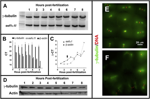
Maternal-zygotic zebrafish ?-tubulin expression and subcellular localization during early development. A. RT-PCR analysis revealed by electrophoresis in agarose gel showed that ?-tubulin mRNA is detected during the first 8?h of the zebrafish embryogenesis. B. qPCR analysis indicated that ?-tubulin mRNA expression levels were invariant during the first 8?h of zebrafish embryogenesis. ?-actin and eef1a were used as reference genes. C. Immunoblot analysis showed that ?-tubulin protein is present during the cleavage (1?3?hpf) and post-MBT stages (3?8?hpf) of zebrafish development. Actin was used as a loading control. D?E. Fluorescence microscopy images of zebrafish embryos (2?hpf) double-stained with anti-?-tubulin antibody (green) and DAPI (red). Metaphase (E), anaphase, and telophase (F) stages of the cell cycle are shown. |
| Gene: | |
|---|---|
| Antibody: | |
| Fish: | |
| Anatomical Terms: | |
| Stage Range: | 4-cell to 75%-epiboly |
Reprinted from Mechanisms of Development, 154, Pouchucq, L., Undurraga, C.A., Fuentes, R., Cornejo, M., Allende, M.L., Monasterio, O., γ-Tubulin small complex formation is essential for early zebrafish embryogenesis, 145-152, Copyright (2018) with permission from Elsevier. Full text @ Mech. Dev.