FIGURE

Fig. 7

ID
ZDB-FIG-181213-12
Publication
Gurung et al., 2018 - Distinct roles for the cell adhesion molecule Contactin2 in the development and function of neural circuits in zebrafish
Other Figures
All Figure Page
Back to All Figure Page
Fig. 7

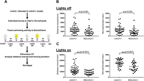
cntn2 mutants exhibit swimming deficits.

(A) Swimming assay and analysis. (B, C) Distance moved and moving duration are compared between cntn2+/− heterozygote and MZcntn2−/− mutant larvae during Lights off (B) and Lights on (C) phases. There were significant differences between cntn2+/− (n = 43) and MZcntn2−/− (n = 27). MZcntn2−/− larvae moved less than cntn2+/− siblings during both lights off (unpaired t-test, *p < 0.05) and lights on (unpaired t-test with Welch's correction, **p < 0.001) phases (B, C). MZcntn2−/− larvae also moved for shorter duration compared to cntn2+/− siblings during both lights off (unpaired t-test, *p < 0.05) and lights on (unpaired t-test, **p < 0.001) phases (B, C). Error bars show Mean ± SD.

Expression Data

Expression Detail
Antibody Labeling
Phenotype Data
Fish:
Observed In:
Stage: Days 7-13

Phenotype Detail
Acknowledgments
This image is the copyrighted work of the attributed author or publisher, and ZFIN has permission only to display this image to its users. Additional permissions should be obtained from the applicable author or publisher of the image.

Reprinted from Mechanisms of Development, 152, Gurung, S., Asante, E., Hummel, D., Williams, A., Feldman-Schultz, O., Halloran, M.C., Sittaramane, V., Chandrasekhar, A., Distinct roles for the cell adhesion molecule Contactin2 in the development and function of neural circuits in zebrafish, 1-12, Copyright (2018) with permission from Elsevier. Full text @ Mech. Dev.