FIGURE
Fig. S4
- ID
- ZDB-FIG-180912-10
- Publication
- Gurung et al., 2018 - Distinct roles for the cell adhesion molecule Contactin2 in the development and function of neural circuits in zebrafish
- Other Figures
- All Figure Page
- Back to All Figure Page
Fig. S4
|
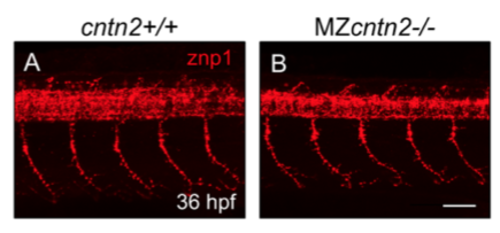
Spinal motor axons develop normally in cntn2 mutants Lateral views of the trunk with anterior to the left, in 36 hpf embryos stained with znp1 antibody. (A) In a wildtype embryo, the motor axon fascicles have extended into the ventral trunk musculature. (B) In a MZcntn2 mutant, the pattern of motor axon outgrowth is not affected. Scale bar in B, 50 ?m for A and B. |
Expression Data
Expression Detail
Antibody Labeling
Phenotype Data
Phenotype Detail
Acknowledgments
This image is the copyrighted work of the attributed author or publisher, and
ZFIN has permission only to display this image to its users.
Additional permissions should be obtained from the applicable author or publisher of the image.
Reprinted from Mechanisms of Development, 152, Gurung, S., Asante, E., Hummel, D., Williams, A., Feldman-Schultz, O., Halloran, M.C., Sittaramane, V., Chandrasekhar, A., Distinct roles for the cell adhesion molecule Contactin2 in the development and function of neural circuits in zebrafish, 1-12, Copyright (2018) with permission from Elsevier. Full text @ Mech. Dev.

